Review
. 2020 Jul 1;100(3):945-982.
doi: 10.1152/physrev.00017.2019. Epub 2019 Dec 23.

Physiology and Pathophysiology of Itch

Affiliations
Review

Physiology and Pathophysiology of Itch

Ferda Cevikbas et al. Physiol Rev. .

Abstract

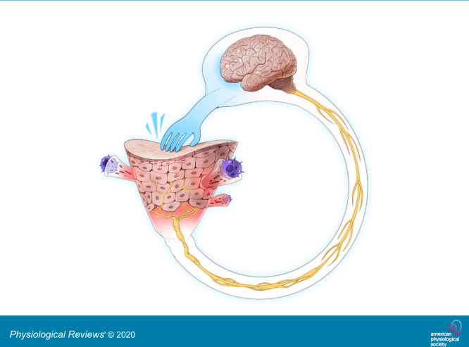
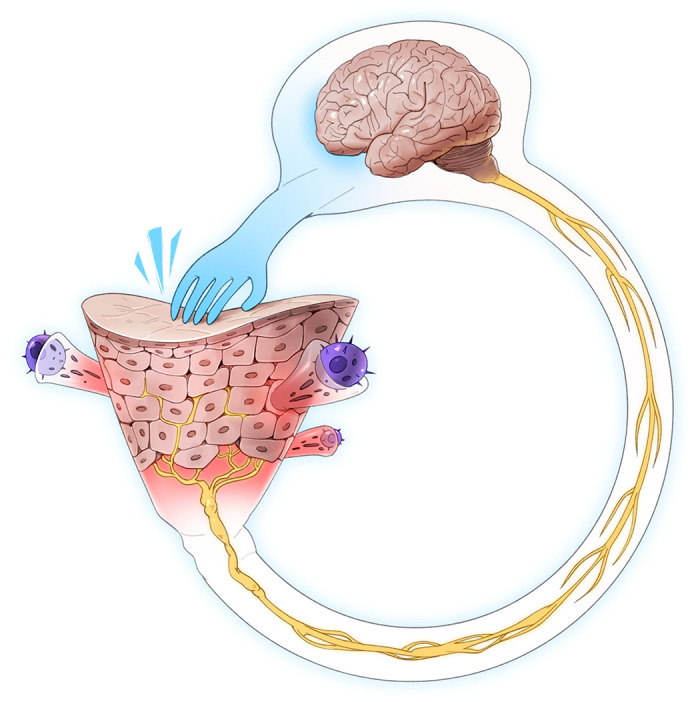
Itch is a topic to which everyone can relate. The physiological roles of itch are increasingly understood and appreciated. The pathophysiological consequences of itch impact quality of life as much as pain. These dynamics have led to increasingly deep dives into the mechanisms that underlie and contribute to the sensation of itch. When the prior review on the physiology of itching was published in this journal in 1941, itch was a black box of interest to a small number of neuroscientists and dermatologists. Itch is now appreciated as a complex and colorful Rubik's cube. Acute and chronic itch are being carefully scratched apart and reassembled by puzzle solvers across the biomedical spectrum. New mediators are being identified. Mechanisms blur boundaries of the circuitry that blend neuroscience and immunology. Measures involve psychophysics and behavioral psychology. The efforts associated with these approaches are positively impacting the care of itchy patients. There is now the potential to markedly alleviate chronic itch, a condition that does not end life, but often ruins it. We review the itch field and provide a current understanding of the pathophysiology of itch. Itch is a disease, not only a symptom of disease.

Keywords: atopic dermatitis; itch; neurogenic inflammation; pruritus; quality of life.


Conflict of interest statement

F. Cevikbas holds equity in Dermira, Inc. E. Lerner is on the Scientific Advisory Board of Escient Pharmaceuticals.

Figures

None
Graphical abstract
FIGURE 1.
FIGURE 1.
Initiation of itch. Allergens, pruritogens, and irritants are exogenous substances which interface with the skin in acute and chronic itch. Branching terminal fibers of afferent neurons which sense these substances reach the epidermis, the uppermost viable layer of skin immediately below the stratum corneum barrier. The sensory neurons are considered histaminergic or non-histaminergic. Neural activity drives the recruitment of immune cells, including mast cells and CD4+ T cells among others. These cells release mediators that activate cognate receptors on sensory neurons to release neuropeptides to contribute to the itch-scratch cycle. Messages are relayed from the peripheral afferents to their cell bodies in dorsal root or trigeminal ganglia followed by synapsing with second-order neurons in the spinal cord. The thalamus then assists in the interpretation of messages encoding itch. IL, interleukin; STT, spinothalamic tract.
FIGURE 2.
FIGURE 2.
Itch circuitry from the periphery to the brain. Itch-related G protein-coupled receptors (GPCRs) and cytokine receptors on sensory neurons from the skin to the spinal cord are categorized as NP1, NP2, and NP3 neurons. Some receptors are expressed in all NP1–3 neurons, whereas MrgprA3, an itch specific marker, is present only in the NP2 population. The natriuretic peptide Nppb acts on its receptor NPR1 to link transmission between dorsal root ganglion (DRG) and spinal neurons. Complex inhibitory and excitatory interneuron activity modulates the signal that will be projected to the brain. Gastrin releasing peptide (GRP) and GRP receptor (GRPR) are each key to excitatory itch circuits in the spinal cord. Inhibitory basic helix loop helix 5 interneurons (B5-I) and somatostatin (SOM) interneurons have inhibitory function. Loss of inhibition either by neuronal depletion of B5-I interneurons or the inhibitory neurotransmitters GABA, glycine, or dynorphin results in intensified itch. This pattern most likely occurs in chronic itch conditions. Enkephalinergic interneurons are important in the gate control between intense pain and itch sensation. Mechanical itch is transmitted through a different, so far unclassified, neuron population and second-order neurons in the spinal cord. The circuitry does not involve excitatory GRP but includes the inhibitory neuropeptide NPY (neuropeptide Y). Contagious itch in mice is relayed through GRPR+ neurons in the suprachiasmatic nucleus. [Modified from Dong and Dong (71), with permission from Elsevier.]
FIGURE 3.
FIGURE 3.
Glia-neuron interaction. A: in the periphery, Schwann cells are thought to modulate itch via TRPA1 channels and the release of CCL2. While expressed in neurons and Schwann glial cells (SGC), silencing of TRPA1 in the latter resulted in reduction of sensitization and signs of inflammation. B: spinal cord glia comprises microglia, astrocytes, and oligodendrocytes. Pruriceptors, a subpopulation of nociceptors, target lamina II inner (IIi). Activated microglia as detected by increased p38 mitogen-activated protein kinase (MAPK) signaling (323) might modulate the pruriceptors and the gastrin releasing peptide receptor (GRPR) neurons (itch signaling second-order neurons). The close proximity of GRPR and gastrin releasing peptide (GRP) in spinal cord neurons enables direct interaction. Itch signaling and amplification of the itch signal might result in a decrease of the inhibitory neurotransmitters (GABA, dynorphin, glycine) and simultaneously increase the excitatory activity via GRP. Activated astrocytes (astrogliosis), detected by increased signal transducers and activators of transcription 3 (STAT3) immunolabeling, release lipocalin 2 (LCN2) which plays a major role in itch progression and maintenance of chronic itch.
FIGURE 4.
FIGURE 4.
Mrgpr gene loci and receptor ligands in mice and humans. All Mrgpr genes are located on chromosome 7 in the mouse and chromosome 11 in humans. While still considered orphan receptors, substances known to activate specific mouse and human Mrgprs are indicated. Note that some substances which activate a single human receptor interact with more than one mouse Mrgpr. Similarly, some substances which activate a single mouse Mrgpr interact with more than one human receptor.
FIGURE 5.
FIGURE 5.
Sensory neuronal receptors and signaling molecules for the transduction of itch. Sensory afferents express a multitude of receptors, providing redundancy to ensure the transmission of acute and chronic itch signaling. A number of these receptors are depicted here, including serotonin-5-hydroxytryptamine receptors (5-HTRs), promiscuous murine itch receptors MrgprA1/A3/C11, cytokine signaling receptor complexes interleukin (IL)-31RA, oncostatin M receptor β (OSMRβ), and IL-4Ra and IL-13R and voltage-gated sodium channels (Nav). A plethora of downstream signaling molecules link receptor activation with generation of action potentials, the details of which remain to be determined. In persistent itch, signaling might increase receptor expression, including the itch-related signaling transducer channels TRPV1 and TRPA1. It remains to be investigated whether downstream signaling mediators such as the mitogen-activated protein kinase (MAPK) and Janus activated kinase (JAK)/signal transducers and activators of transcription (STAT) acutely alter itch-relevant receptors via sensitization or on transcriptional levels. BAM8-22, bovine adrenal medulla 8-22; PKC, protein kinase C; PLA, phospholipase A; PLC, phospholipase C; SP, substance P.
FIGURE 6.
FIGURE 6.
Acute and chronic itch in humans. In acute itch, histaminergic itch is induced by histamine via activation of its receptors on sensory neurons. Non-histaminergic itch can be induced by a plethora of molecules, of which some are identified. One such molecule is interleukin (IL)-31 which is released from immune cells including CD4+ TH2 cells and mast cells. Upon release, IL-31 activates a heterodimeric receptor complex consisting of IL-31Rα and oncostatin M receptor β (OSMRβ) on sensory neurons to elicit itch directly. It is possible that this process induces the release of neuropeptides such as substance P (SP) from sensory neurons. SP activates MRGPRX2 on mast cells and may be induced on sensory neurons. Itch triggers degranulation of mast cells and the differential and controlled liberation of various neuropeptides, proteases, and cytokines. A continuous inflammatory milieu may result in chronic itch, which is debilitating in many diseases. IL-31 has been linked to chronic itch skin conditions. During inflammation, immune cells release other cytokines including IL-4 and IL-13 which signal via the heterodimer IL-4Rα and IL-13Rα1 and are reported to function as neuronal enhancers for different itch pathways. Bile acids have recently been reported to activate MRGPRX4 and may thus contribute to cholestatic itch.
FIGURE 7.
FIGURE 7.
Peripheral and central targets of itch. Peripheral itch targets are receptors and mediators of chronic itch including the interleukin (IL)-31 pathway. Targeting IL-4 and IL-13 with dupilumab has been successful in atopic dermatitis. Antagonists of MRGPRX2 might be beneficial in the treatment of inflammation, itch, and urticaria. Antagonism of MRGPX4 has the potential to benefit the treatment of cholestatic itch when driven by bile acids. In neuropathic pain, targeting peripheral GABA-R has proven analgesic properties, which could be translated to itch. Central targets of itch act in the central nervous system and include the use of κ-opioid receptor (KOR) agonists and μ-opioid receptor (MOR) antagonists. Targeting spinal serotonin-pathways via 5-hydroxytryptamine (5-HT) 3 and 7 receptors may achieve clinical success in chronic itch management. Antagonists directed against the NK1R most likely work spinally. Targeting itch-specific circuits such as gastrin releasing peptide receptor (GRPR) might represent a therapeutic avenue. Increasing inhibitory tone via replenishing GABA reduces acute and chronic itch in mice. Gabapentin and pregabalin, widely used to treat neuropathic pain, are prescribed in the clinic to treat chronic itch patients. DRG, dorsal root ganglion; GRP, gastrin releasing peptide; SP, substance P.
FIGURE 8.
FIGURE 8.
Itch-scratch cycle. The many areas where therapeutic approaches can be directed are depicted. These areas range from the skin barrier, depicted as a funnel with associated immune cells together with afferent fibers that flow into the spinal circuitry and on to the brain.

References

    1. Abdo H, Calvo-Enrique L, Lopez JM, Song J, Zhang M-D, Usoskin D, El Manira A, Adameyko I, Hjerling-Leffler J, Ernfors P. Specialized cutaneous Schwann cells initiate pain sensation. Science 365: 695–699, 2019. doi: 10.1126/science.aax6452. - DOI - PubMed
    1. Akdis CA, Simons FER. Histamine receptors are hot in immunopharmacology. Eur J Pharmacol 533: 69–76, 2006. doi: 10.1016/j.ejphar.2005.12.044. - DOI - PubMed
    1. Akiyama T, Carstens MI, Carstens E. Enhanced scratching evoked by PAR-2 agonist and 5-HT but not histamine in a mouse model of chronic dry skin itch. Pain 151: 378–383, 2010. doi: 10.1016/j.pain.2010.07.024. - DOI - PMC - PubMed
    1. Akiyama T, Carstens MI, Carstens E. Differential itch- and pain-related behavioral responses and µ-opoid modulation in mice. Acta Derm Venereol 90: 575–581, 2010. doi: 10.2340/00015555-0962. - DOI - PubMed
    1. Akiyama T, Carstens MI, Ikoma A, Cevikbas F, Steinhoff M, Carstens E. Mouse model of touch-evoked itch (alloknesis). J Invest Dermatol 132: 1886–1891, 2012. doi: 10.1038/jid.2012.52. - DOI - PMC - PubMed

Publication types