Resolving homology in the face of shifting germ layer origins: Lessons from a major skull vault boundary
- PMID: 31869306
- PMCID: PMC6927740
- DOI: 10.7554/eLife.52814
Resolving homology in the face of shifting germ layer origins: Lessons from a major skull vault boundary
Abstract
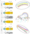
The vertebrate skull varies widely in shape, accommodating diverse strategies of feeding and predation. The braincase is composed of several flat bones that meet at flexible joints called sutures. Nearly all vertebrates have a prominent 'coronal' suture that separates the front and back of the skull. This suture can develop entirely within mesoderm-derived tissue, neural crest-derived tissue, or at the boundary of the two. Recent paleontological findings and genetic insights in non-mammalian model organisms serve to revise fundamental knowledge on the development and evolution of this suture. Growing evidence supports a decoupling of the germ layer origins of the mesenchyme that forms the calvarial bones from inductive signaling that establishes discrete bone centers. Changes in these relationships facilitate skull evolution and may create susceptibility to disease. These concepts provide a general framework for approaching issues of homology in cases where germ layer origins have shifted during evolution.
Keywords: developmental biology; evolutionary biology; homology; skull; sutures.
© 2019, Teng et al.
Conflict of interest statement
CT, LC, RM, MS, JC No competing interests declared
Figures


Similar articles
-
Tissue origins and interactions in the mammalian skull vault.Dev Biol. 2002 Jan 1;241(1):106-16. doi: 10.1006/dbio.2001.0487. Dev Biol. 2002. PMID: 11784098
-
The Development of the Calvarial Bones and Sutures and the Pathophysiology of Craniosynostosis.Curr Top Dev Biol. 2015;115:131-56. doi: 10.1016/bs.ctdb.2015.07.004. Epub 2015 Oct 1. Curr Top Dev Biol. 2015. PMID: 26589924 Review.
-
Vertebrate cranial mesoderm: developmental trajectory and evolutionary origin.Cell Mol Life Sci. 2020 May;77(10):1933-1945. doi: 10.1007/s00018-019-03373-1. Epub 2019 Nov 13. Cell Mol Life Sci. 2020. PMID: 31722070 Free PMC article. Review.
-
Developmental anatomy of craniofacial sutures.Front Oral Biol. 2008;12:1-21. doi: 10.1159/000115028. Front Oral Biol. 2008. PMID: 18391492 Review.
-
Cranial suture lineage and contributions to repair of the mouse skull.Development. 2024 Feb 1;151(3):dev202116. doi: 10.1242/dev.202116. Epub 2024 Feb 12. Development. 2024. PMID: 38345329 Free PMC article.
Cited by
-
From head to tail: regionalization of the neural crest.Development. 2020 Oct 26;147(20):dev193888. doi: 10.1242/dev.193888. Development. 2020. PMID: 33106325 Free PMC article. Review.
-
Modularity, homology, heterochrony: Gavin de Beer's legacy to the mammalian skull.Philos Trans R Soc Lond B Biol Sci. 2023 Jul 3;378(1880):20220078. doi: 10.1098/rstb.2022.0078. Epub 2023 May 15. Philos Trans R Soc Lond B Biol Sci. 2023. PMID: 37183898 Free PMC article. Review.
-
A new exceptionally well-preserved basal actinopterygian fish in the juvenile stage from the Upper Triassic Amisan Formation of South Korea.Sci Rep. 2024 Jan 3;14(1):317. doi: 10.1038/s41598-023-50803-z. Sci Rep. 2024. PMID: 38172381 Free PMC article.
-
Making and shaping endochondral and intramembranous bones.Dev Dyn. 2021 Mar;250(3):414-449. doi: 10.1002/dvdy.278. Epub 2020 Dec 28. Dev Dyn. 2021. PMID: 33314394 Free PMC article. Review.
-
Developmental fates of shark head cavities reveal mesodermal contributions to tendon progenitor cells in extraocular muscles.Zoological Lett. 2021 Feb 15;7(1):3. doi: 10.1186/s40851-021-00170-2. Zoological Lett. 2021. PMID: 33588955 Free PMC article.
References
-
- Arratia G, Cloutier R. Reassessment of the morphology of Cheirolepis canadensis (Actinopterygii) In: Schultze H-P, Cloutier R, editors. Devonian Fishes and Plants of Miguasha. Quebec, Canada: Taylor & Francis, Ltd; 1996. pp. 165–197.
-
- Benton MJ, Donoghue PCJ, Vinther J, Asher RJ, Friedman M, Near TJ. Constraints on the timescale of animal evolutionary history. Palaeontologia Electronica. 2015:18.1.1FC. doi: 10.26879/424. - DOI
Publication types
MeSH terms
Grants and funding
LinkOut - more resources
Full Text Sources

