. 2019 Dec 26;116(52):26305-26312.
doi: 10.1073/pnas.1902293116. Epub 2019 Dec 23.

The contribution of nonhuman primate research to the understanding of emotion and cognition and its clinical relevance

Affiliations

The contribution of nonhuman primate research to the understanding of emotion and cognition and its clinical relevance

Silvia Bernardi et al. Proc Natl Acad Sci U S A. .

Abstract

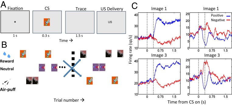
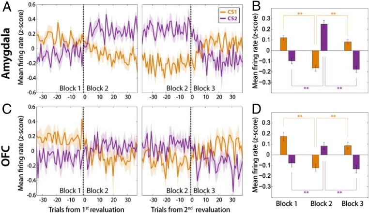
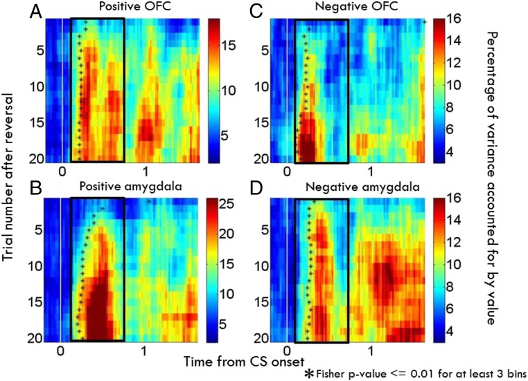
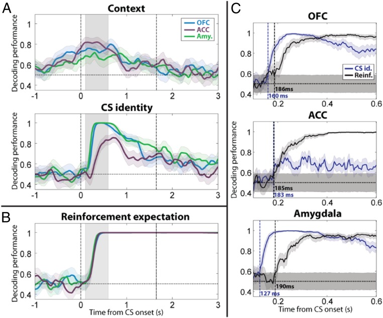
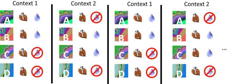
Psychiatric disorders are often conceptualized as arising from dysfunctional interactions between neural systems mediating cognitive and emotional processes. Mechanistic insights into these interactions have been lacking in part because most work in emotions has occurred in rodents, often without concurrent manipulations of cognitive variables. Nonhuman primate (NHP) model systems provide a powerful platform for investigating interactions between cognitive operations and emotions due to NHPs' strong homology with humans in behavioral repertoire and brain anatomy. Recent electrophysiological studies in NHPs have delineated how neural signals in the amygdala, a brain structure linked to emotion, predict impending appetitive and aversive stimuli. In addition, abstract conceptual information has also been shown to be represented in the amygdala and in interconnected brain structures such as the hippocampus and prefrontal cortex. Flexible adjustments of emotional behavior require the ability to apply conceptual knowledge and generalize to different, often novel, situations, a hallmark example of interactions between cognitive and emotional processes. Elucidating the neural mechanisms that explain how the brain processes conceptual information in relation to emotional variables promises to provide important insights into the pathophysiology accounting for symptoms in neuropsychiatric disorders.

Keywords: abstraction; amygdala; emotion; nonhuman primates; prefrontal cortex.


Conflict of interest statement

The authors declare no competing interest.

Figures

Fig. 1.
Fig. 1.
Amygdala neurons represent the positive and negative valence of CSs. (A) Sequence of a trace-conditioning trial. Monkeys centered gaze at a fixation point for 1 s and viewed a fractal image for 300 ms. US delivery followed a 1,500-ms trace interval. (B) Task structure. Positive images, liquid reward; negative images, aversive air puff; nonreinforced images, no US. After monkeys learned initial contingencies, image reinforcement contingencies switched without warning. (C) PSTHs for 2 neurons. Reward trials, blue; air puff trials, red. Image 1 was initially rewarded, then paired with air puff after reversal; image 3, opposite contingencies. (Left column) Positive value-coding neuron, responding more strongly to both images when rewarded. (Right column) Negative value-coding neuron, responding most strongly when air puff follows each image presentation. Reprinted with permission from ref. .
Fig. 2.
Fig. 2.
Neurons in the amygdala and OFC represent the relative amount of reward associated with a CS. (A) Population average firing rate plotted as a function of trial number for amygdala neurons that responded selectively to the amount of expected reward. (Left) Activity changes in relation to a revaluation of CS1 (orange), which occurs by increasing the reward amount associated with CS2 (purple). (Right) Activity changes in relation to a second revaluation of CS1, which now occurs by decreasing the reward amount associated with CS2. (B) Average firing rate of neurons in A for each block. **P < 0.01 (Wilcoxon sign-rank test). (C and D) Same as A and B, except for neurons recorded in OFC. Adapted from ref. , with permission from Elsevier.
Fig. 3.
Fig. 3.
Neurons within appetitive and aversive networks in amygdala and OFC update responses to changed reinforcement contingencies at different rates. Time course of changes in value-related signals in amygdala and OFC plotted as a function of time and trial number relative to reversal (AD). For each bin, an index computed for each cell the proportion of variance accounted for by image value divided by the total variance using a 2-way ANOVA (61), and this index was averaged across populations. (A and B) Average contribution of image value in positive value-coding neurons in OFC (A) and amygdala (B). (C and D) Same as A and B, except for negative value-coding cells. Black asterisks, time when the contribution-of-value index becomes significant (asterisks placed in center of the first of at least 3 consecutive significant bins; Fisher P < 0.01). Bin size, 200 ms. Bin steps, 20 ms. Adapted from ref. , with permission from Elsevier.
Fig. 4.
Fig. 4.
Neural ensembles in amygdala, OFC, and ACC encode uncued contexts, stimulus identity, and expected reinforcement. (A) Decoding performance for context and CS identity in amygdala, OFC, and ACC (250-ms sliding window, 50-ms steps). Blue, OFC; purple, ACC; green, amygdala. Shaded areas, 95% confidence intervals (bootstrap). Vertical dashed lines, CS onset and earliest possible US onset. (B) Decoding performance for reinforcement expectation plotted vs. time relative to image onset. (C) Timing of onset of CS identity (blue) and reinforcement expectation (black) signals in OFC, ACC, and amygdala (50-ms sliding window, 5-ms steps for 500-ms window shown in A and B by gray shading). Vertical dashed lines (and labels) indicate the first time bin where decoding performance is significantly above chance level and remains there for 10 time bins. Shaded areas, 95% confidence intervals around chance (shuffle). Adapted from ref. , with permission from Elsevier.
Fig. 5.
Fig. 5.
Serial reversal-learning task in which 2 stimulus–response–outcome task sets define 2 contexts. Monkeys hold a bar and then fixate to begin a trial. Upon viewing an image, monkeys must hold or release a bar to perform correctly. Two stimuli in each context were rewarded after correct decisions (context 1: A, C; context 2: B, C).

References

    1. Ongür D., Price J. L., The organization of networks within the orbital and medial prefrontal cortex of rats, monkeys and humans. Cereb. Cortex 10, 206–219 (2000). - PubMed
    1. Petrides M., Pandya D. N., Dorsolateral prefrontal cortex: Comparative cytoarchitectonic analysis in the human and the macaque brain and corticocortical connection patterns. Eur. J. Neurosci. 11, 1011–1036 (1999). - PubMed
    1. Petrides M., Pandya D. N., Comparative cytoarchitectonic analysis of the human and the macaque ventrolateral prefrontal cortex and corticocortical connection patterns in the monkey. Eur. J. Neurosci. 16, 291–310 (2002). - PubMed
    1. Bouret S., Richmond B. J., Ventromedial and orbital prefrontal neurons differentially encode internally and externally driven motivational values in monkeys. J. Neurosci. 30, 8591–8601 (2010). - PMC - PubMed
    1. Cai X., Padoa-Schioppa C., Contributions of orbitofrontal and lateral prefrontal cortices to economic choice and the good-to-action transformation. Neuron 81, 1140–1151 (2014). - PMC - PubMed

LinkOut - more resources