. 2020 Jan 7;117(1):214-220.
doi: 10.1073/pnas.1910343117. Epub 2019 Dec 23.

Biodegradable nanofiber-based piezoelectric transducer

Affiliations

Biodegradable nanofiber-based piezoelectric transducer

Eli J Curry et al. Proc Natl Acad Sci U S A. .

Abstract

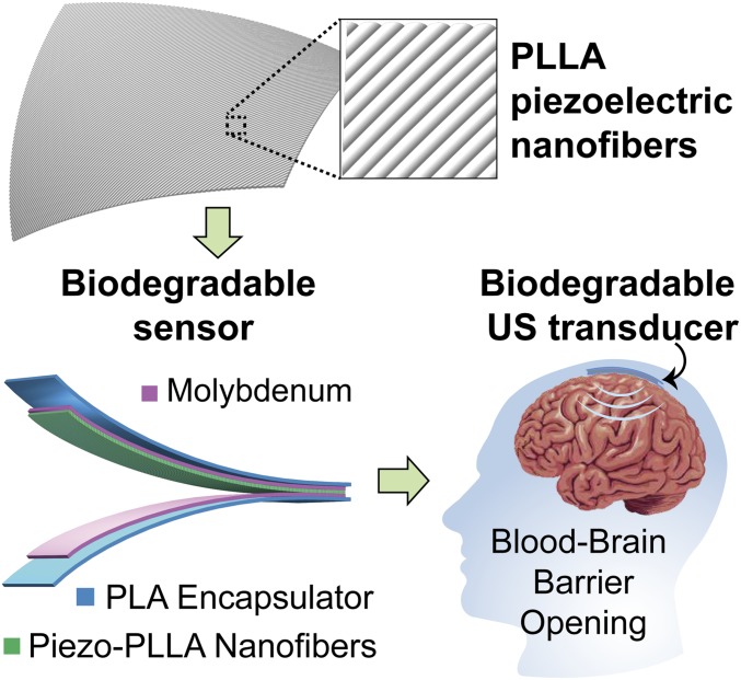
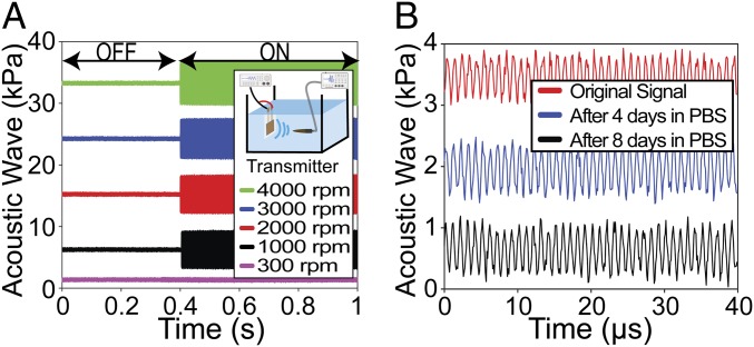
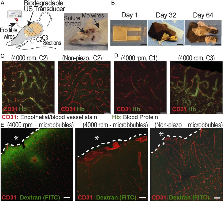
Piezoelectric materials, a type of "smart" material that generates electricity while deforming and vice versa, have been used extensively for many important implantable medical devices such as sensors, transducers, and actuators. However, commonly utilized piezoelectric materials are either toxic or nondegradable. Thus, implanted devices employing these materials raise a significant concern in terms of safety issues and often require an invasive removal surgery, which can damage directly interfaced tissues/organs. Here, we present a strategy for materials processing, device assembly, and electronic integration to 1) create biodegradable and biocompatible piezoelectric PLLA [poly(l-lactic acid)] nanofibers with a highly controllable, efficient, and stable piezoelectric performance, and 2) demonstrate device applications of this nanomaterial, including a highly sensitive biodegradable pressure sensor for monitoring vital physiological pressures and a biodegradable ultrasonic transducer for blood-brain barrier opening that can be used to facilitate the delivery of drugs into the brain. These significant applications, which have not been achieved so far by conventional piezoelectric materials and bulk piezoelectric PLLA, demonstrate the PLLA nanofibers as a powerful material platform that offers a profound impact on various medical fields including drug delivery, tissue engineering, and implanted medical devices.

Keywords: PLLA; biodegradable; piezoelectric; pressure sensors; ultrasound transducer.


Conflict of interest statement

The authors declare no competing interest.

Figures

Fig. 1.
Fig. 1.
PLLA nanofibers with highly controllable and excellent piezoelectric performance for biodegradable implanted piezoelectric devices. The image at Top is a simplified schematic of the treated piezoelectric PLLA nanofibers. The image at Bottom Left is the schematic of a biodegradable pressure sensor and ultrasound (US) transducer. The image at Bottom Right is a schematic illustrating the biodegradable US transducer, implanted inside the brain, which can repeatedly induce US to open the blood–brain barrier (BBB) and facilitate the delivery of drugs into the brain.
Fig. 2.
Fig. 2.
Material characterization of the electrospun PLLA. (A) Results from differential scanning calorimetry (DSC) of electrospun PLLA nanofiber films collected at different spin speeds. (B) The 2D X-ray diffraction (2D XRD) images show orientation of crystal domains inside the electrospun PLLA nanofibers, made with different collection speeds. (C) Scanning electron microscopy (SEM) images show PLLA nanofiber alignment with different collection speeds. (Scale bars, 40 µm.) (D) Graphical summary illustrating the trend that, as the PLLA nanofibers are collected at faster speeds, the Herman orientation factor (i.e., crystal alignment) and crystallinity percentage generally increase.
Fig. 3.
Fig. 3.
Piezoelectric characterization of the treated PLLA nanofiber films. (A) Charge output from stretched, bulk piezo-PLLA (yellow) and treated electrospun PLLA, collected at different speeds, under the same impact force. (B) Displacement of stretched, bulk PLLA (yellow) and treated electrospun PLLA, collected at different speeds, under the same voltage (20 Vpp) at 1 Hz. (C) Displacement of 300 rpm PLLA negative-control sample (red) and 4,000 rpm PLLA (black), under increasing magnitudes of voltage at 1 Hz. (D) Comparison of the piezoelectric performance for 3,000 rpm electrospun PLLA samples annealed under different conditions over a 14-d period.
Fig. 4.
Fig. 4.
Wireless, biodegradable PLLA-nanofiber force sensor. (A) Comparison of calibration curves for a biodegradable sensor using stretched, bulk piezo-PLLA film (black) and a 4,000 rpm electrospun PLLA nanofiber film (red). Inset shows the optical image of the biodegradable and flexible force sensor, made from the PLLA nanofibers. (Scale bar, 5 mm.) (B) Output from a charge amplifying circuit connected to a 4,000 rpm electrospun, biodegradable PLLA sensor that is subjected to 10,000 cycles of a 10-N force. (C) Simplified schematic of the implanted, wireless pressure sensor in a mouse (Left) and optical image of a mouse receiving the wireless PLLA sensor implanted (Right). NFC, near-field communication chip. (D) Comparison of the simulated abdominal pressure signals, wirelessly recorded from an implanted biodegradable PLLA nanofiber sensor using a 300 rpm negative control (black) and a 4,000 rpm film (red).
Fig. 5.
Fig. 5.
US characterization of the biodegradable PLLA-nanofiber transducer. (A) The output pressure from the transducer with different electrospinning speeds under the same input voltage. The Inset is the simplified schematic of the experiment. (B) Output pressure from a biodegradable US transducer made from 4,000 rpm electrospun PLLA under the same input voltage after different days in PBS at 37 °C.
Fig. 6.
Fig. 6.
In vivo experiment to demonstrate the application of PLLA nanofiber transducer for the BBB opening and drug delivering. (A) The schematic (Left) and optical image (Right) of the in vivo experiment. (B) The optical images of a typical biodegradable US transducer at different days in the buffered solution at an accelerated-degradation temperature of 70 °C. (Scale bars, 5 mm.) (C) Representative images showing the autofluorescent signal of blood protein at the coronal section (C2) from the brains of mice that received US from the 4,000 rpm PLLA transducer (Left) and the 300 rpm PLLA negative-control transducer (Right). (D) Representative images show the blood protein signal at different coronal sections of the same mouse brain receiving the US treatment. Section C3 (Right) is closer to the implanted transducer, while section C1 (Left) is far away from the implanted US transducer, serving as an internal control. (Scale bars in C and D, 30 µm.) (E) Representative images show the signal of dextran (FITC) at the coronal sections from the brains of mice that received different treatments and samples. The dashed lines show the boundary between the brain and the biodegradable device. The asterisk (*) shows the position of the implanted device. (Scale bars, 50 µm.)

References

    1. Chorsi M. T., et al. , Piezoelectric biomaterials for sensors and actuators. Adv. Mater. 31, e1802084 (2019). - PubMed
    1. Nguyen T. D., et al. , Piezoelectric nanoribbons for monitoring cellular deformations. Nat. Nanotechnol. 7, 587–593 (2012). - PubMed
    1. Curry E. J., et al. , Biodegradable piezoelectric force sensor. Proc. Natl. Acad. Sci. U.S.A. 115, 909–914 (2018). - PMC - PubMed
    1. Ikada Y., Shikinami Y., Hara Y., Tagawa M., Fukada E., Enhancement of bone formation by drawn poly(l-lactide). J. Biomed. Mater. Res. 30, 553–558 (1996). - PubMed
    1. Tajitsu Y., Kanesaki M., Tsukiji M., Imoto K., Date M., Fukada E., Novel tweezers for biological cells using piezoelectric polylactic acid fibers. Ferroelectrics 320, 133–139 (2005).

Publication types

LinkOut - more resources