

Mycobacterial HflX is a ribosome splitting factor that mediates antibiotic resistance
- PMID: 31871194
- PMCID: PMC6955381
- DOI: 10.1073/pnas.1906748117
Mycobacterial HflX is a ribosome splitting factor that mediates antibiotic resistance
Abstract
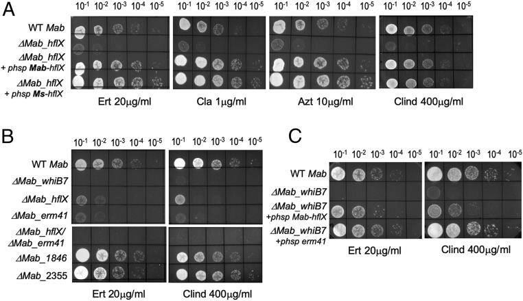
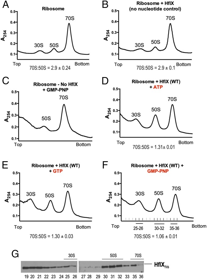
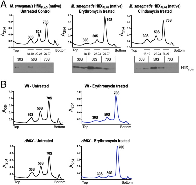
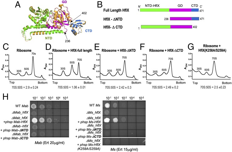
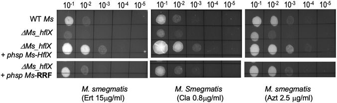
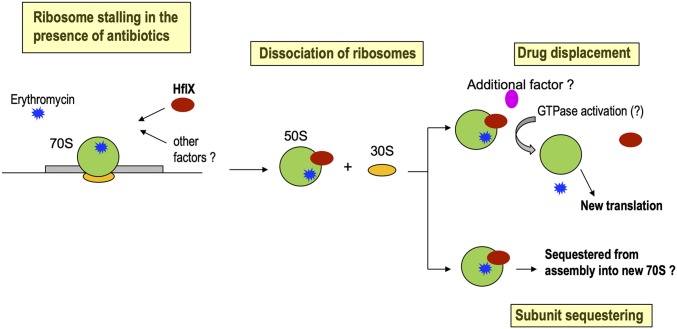
Antibiotic resistance in bacteria is typically conferred by proteins that function as efflux pumps or enzymes that modify either the drug or the antibiotic target. Here we report an unusual mechanism of resistance to macrolide-lincosamide antibiotics mediated by mycobacterial HflX, a conserved ribosome-associated GTPase. We show that deletion of the hflX gene in the pathogenic Mycobacterium abscessus, as well as the nonpathogenic Mycobacterium smegmatis, results in hypersensitivity to the macrolide-lincosamide class of antibiotics. Importantly, the level of resistance provided by Mab_hflX is equivalent to that conferred by erm41, implying that hflX constitutes a significant resistance determinant in M. abscessus We demonstrate that mycobacterial HflX associates with the 50S ribosomal subunits in vivo and can dissociate purified 70S ribosomes in vitro, independent of GTP hydrolysis. The absence of HflX in a ΔMs_hflX strain also results in a significant accumulation of 70S ribosomes upon erythromycin exposure. Finally, a deletion of either the N-terminal or the C-terminal domain of HflX abrogates ribosome splitting and concomitantly abolishes the ability of mutant proteins to mediate antibiotic tolerance. Together, our results suggest a mechanism of macrolide-lincosamide resistance in which the mycobacterial HflX dissociates antibiotic-stalled ribosomes and rescues the bound mRNA. Given the widespread presence of hflX genes, we anticipate this as a generalized mechanism of macrolide resistance used by several bacteria.
Keywords: HflX; Mycobacterium abscessus; erm41; macrolides; ribosome.
Conflict of interest statement
The authors declare no competing interest.
Figures
Similar articles
-
HflX-mediated drug resistance through ribosome splitting and rRNA disordering in mycobacteria.Proc Natl Acad Sci U S A. 2025 Feb 11;122(6):e2419826122. doi: 10.1073/pnas.2419826122. Epub 2025 Feb 6. Proc Natl Acad Sci U S A. 2025. PMID: 39913204 Free PMC article.
-
Ribosome Protection as a Mechanism of Lincosamide Resistance in Mycobacterium abscessus.Antimicrob Agents Chemother. 2021 Oct 18;65(11):e0118421. doi: 10.1128/AAC.01184-21. Epub 2021 Aug 30. Antimicrob Agents Chemother. 2021. PMID: 34460298 Free PMC article.
-
Structural basis for HflXr-mediated antibiotic resistance in Listeria monocytogenes.Nucleic Acids Res. 2022 Oct 28;50(19):11285-11300. doi: 10.1093/nar/gkac934. Nucleic Acids Res. 2022. PMID: 36300626 Free PMC article.
-
Translational attenuation: the regulation of bacterial resistance to the macrolide-lincosamide-streptogramin B antibiotics.CRC Crit Rev Biochem. 1984;16(2):103-32. doi: 10.3109/10409238409102300. CRC Crit Rev Biochem. 1984. PMID: 6203682 Review.
-
Treatment of Mycobacterium abscessus Pulmonary Disease.Chest. 2022 Jan;161(1):64-75. doi: 10.1016/j.chest.2021.07.035. Epub 2021 Jul 24. Chest. 2022. PMID: 34314673 Review.
Cited by
-
Role of a cryptic tRNA gene operon in survival under translational stress.Nucleic Acids Res. 2021 Sep 7;49(15):8757-8776. doi: 10.1093/nar/gkab661. Nucleic Acids Res. 2021. PMID: 34379789 Free PMC article.
-
Unraveling the mechanisms of intrinsic drug resistance in Mycobacterium tuberculosis.Front Cell Infect Microbiol. 2022 Oct 17;12:997283. doi: 10.3389/fcimb.2022.997283. eCollection 2022. Front Cell Infect Microbiol. 2022. PMID: 36325467 Free PMC article. Review.
-
Mechanistic insights into the alternative ribosome recycling by HflXr.Nucleic Acids Res. 2024 Apr 24;52(7):4053-4066. doi: 10.1093/nar/gkae128. Nucleic Acids Res. 2024. PMID: 38407413 Free PMC article.
-
Ribosome hibernation: a new molecular framework for targeting nonreplicating persisters of mycobacteria.Microbiology (Reading). 2021 Feb;167(2):001035. doi: 10.1099/mic.0.001035. Microbiology (Reading). 2021. PMID: 33555244 Free PMC article. Review.
-
Beyond antibiotic resistance: The whiB7 transcription factor coordinates an adaptive response to alanine starvation in mycobacteria.Cell Chem Biol. 2024 Apr 18;31(4):669-682.e7. doi: 10.1016/j.chembiol.2023.12.020. Epub 2024 Jan 23. Cell Chem Biol. 2024. PMID: 38266648 Free PMC article.
References
-
- Griffith D. E., et al. ; ATS Mycobacterial Diseases Subcommittee; American Thoracic Society; Infectious Disease Society of America , An official ATS/IDSA statement: Diagnosis, treatment, and prevention of nontuberculous mycobacterial diseases. Am. J. Respir. Crit. Care Med. 175, 367–416 (2007). Correction in: Am. J. Respir. Crit. Care Med. 175, 744–745 (2007). - PubMed
-
- Nessar R., Cambau E., Reyrat J. M., Murray A., Gicquel B., Mycobacterium abscessus: A new antibiotic nightmare. J. Antimicrob. Chemother. 67, 810–818 (2012). - PubMed
-
- Wilson D. N., The A-Z of bacterial translation inhibitors. Crit. Rev. Biochem. Mol. Biol. 44, 393–433 (2009). - PubMed
Publication types
MeSH terms
Substances
Grants and funding
LinkOut - more resources
Full Text Sources
Medical
Molecular Biology Databases
