. 2019 Dec 26;116(52):26230-26238.
doi: 10.1073/pnas.1903671116. Epub 2019 Dec 23.

Alzheimer's-like pathology in aging rhesus macaques: Unique opportunity to study the etiology and treatment of Alzheimer's disease

Affiliations

Alzheimer's-like pathology in aging rhesus macaques: Unique opportunity to study the etiology and treatment of Alzheimer's disease

Amy F T Arnsten et al. Proc Natl Acad Sci U S A. .

Abstract

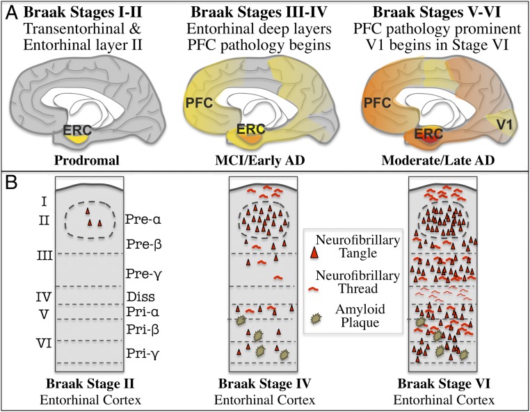
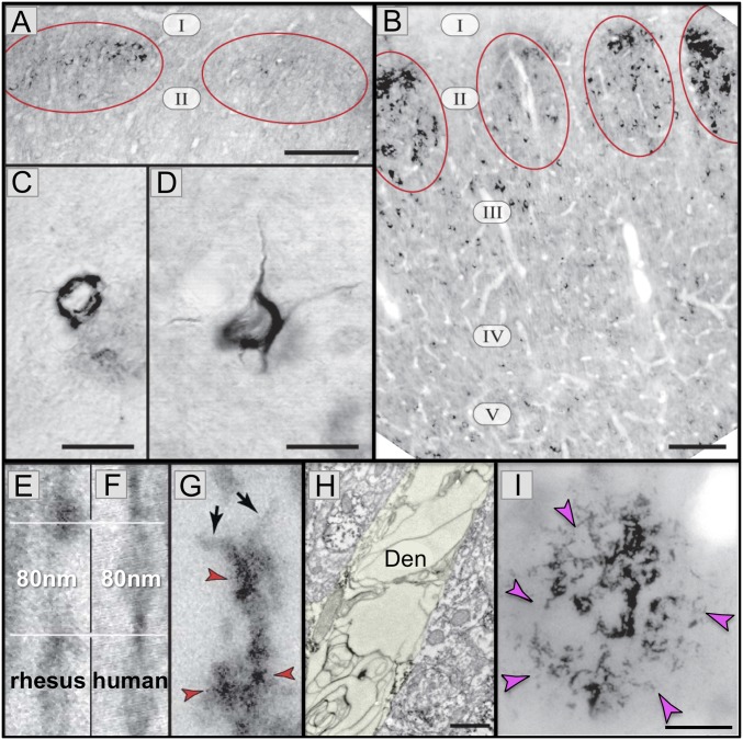
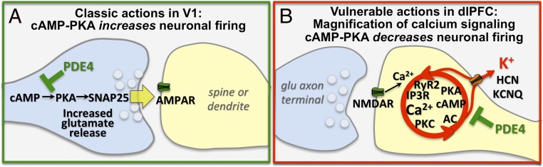
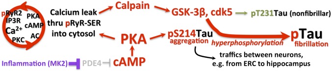
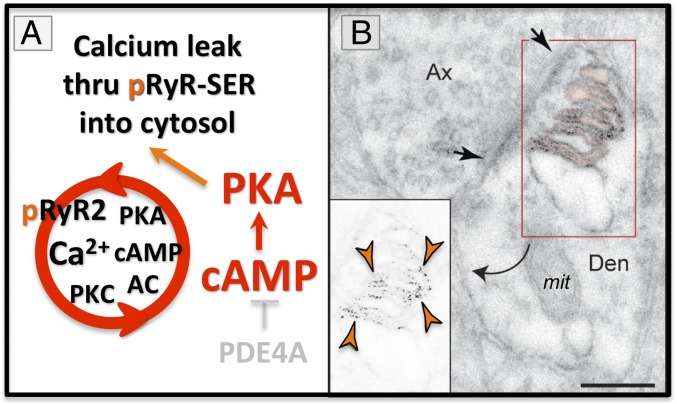
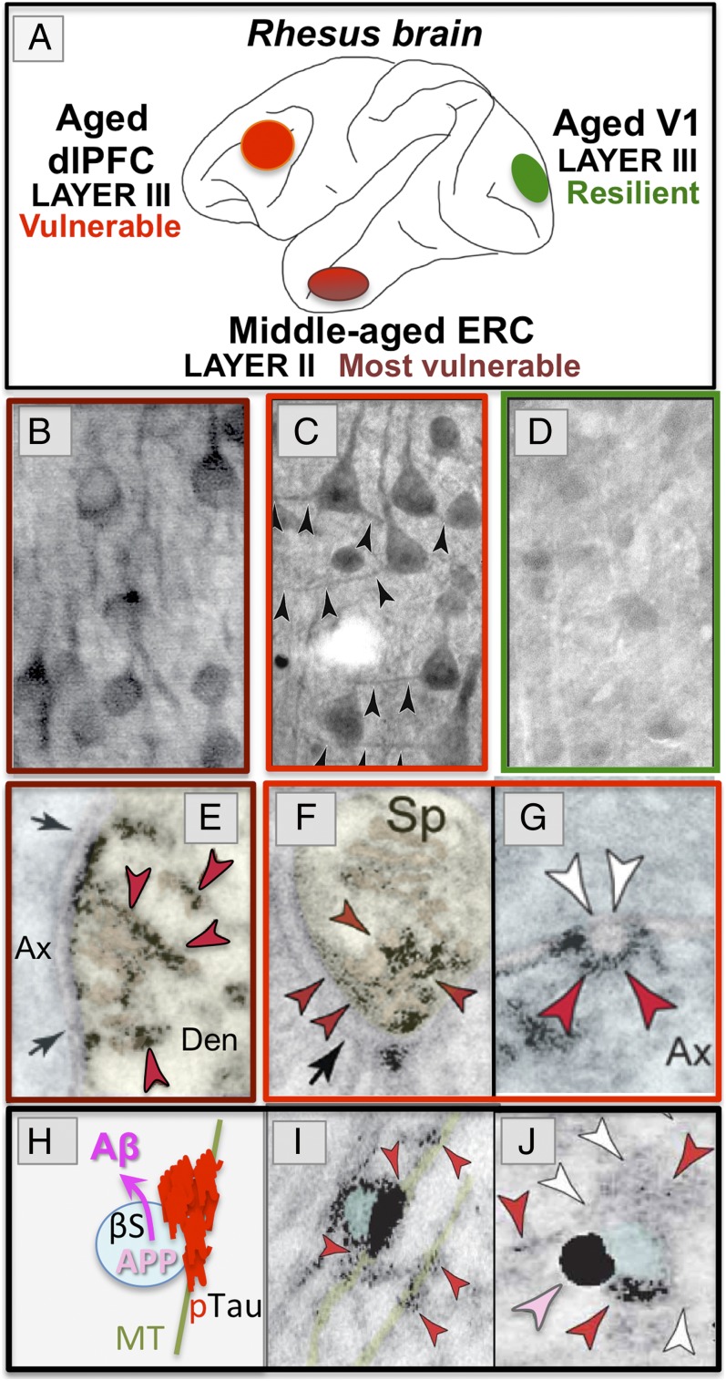
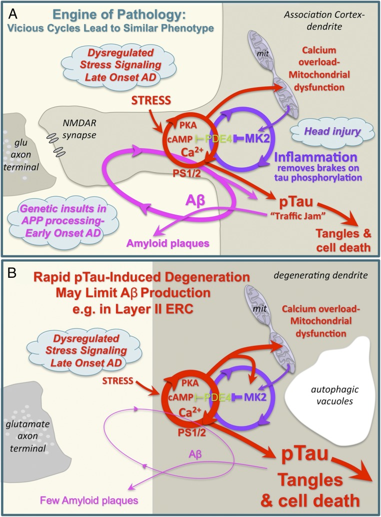
Although mouse models of Alzheimer's disease (AD) have provided tremendous breakthroughs, the etiology of later onset AD remains unknown. In particular, tau pathology in the association cortex is poorly replicated in mouse models. Aging rhesus monkeys naturally develop cognitive deficits, amyloid plaques, and the same qualitative pattern and sequence of tau pathology as humans, with tangles in the oldest animals. Thus, aging rhesus monkeys can play a key role in AD research. For example, aging monkeys can help reveal how synapses in the prefrontal association cortex are uniquely regulated compared to the primary sensory cortex in ways that render them vulnerable to calcium dysregulation and tau phosphorylation, resulting in the selective localization of tau pathology observed in AD. The ability to assay early tau phosphorylation states and perform high-quality immunoelectron microscopy in monkeys is a great advantage, as one can capture early-stage degeneration as it naturally occurs in situ. Our immunoelectron microscopy studies show that phosphorylated tau can induce an "endosomal traffic jam" that drives amyloid precursor protein cleavage to amyloid-β in endosomes. As amyloid-β increases tau phosphorylation, this creates a vicious cycle where varied precipitating factors all lead to a similar phenotype. These data may help explain why circuits with aggressive tau pathology (e.g., entorhinal cortex) may degenerate prior to producing significant amyloid pathology. Aging monkeys therefore can play an important role in identifying and testing potential therapeutics to protect the association cortex, including preventive therapies that are challenging to test in humans.

Keywords: PKA; entorhinal cortex; prefrontal cortex; ryanodine.


Conflict of interest statement

Competing interest statement: A.F.T.A. and Yale University receive royalties from the United States sales of Intuniv by Shire/Takeda Pharmaceuticals. They do not receive royalties from generic or international sales.

Figures

Fig. 1.
Fig. 1.
The stages of Braak tau pathology, showing AT8-labeled, fibrillated tau distribution on a midsagital view of the human brain (A), and in a coronal section of entorhinal cortex (B). Adapted by permission from ref. , Springer Nature: Acta Neuropathologica, copyright 1991.
Fig. 2.
Fig. 2.
AT8 pTau and amyloid pathology in aged rhesus monkeys (26 to 38 y). (A) AT8 label is first seen in layer II cell islands in the ERC in a 26-y-old monkey. (B) In an older monkey (33 y), AT8 labeling is more extensive in layer II and now extends to deeper layers. (C and D) Higher-magnification view of neurofibrillary tangles in layer II (C) and layer V (D) from a 38-y-old monkey. (EG) High-magnification view of AT8-labeled (red arrrowheads) paired helical filaments from an aged monkey (E and G) or human (F). Note the same width (10 nm) and helical frequency (80 nm). Black arrows in G show the abrupt ends of each filament. (H) A degenerating dendrite (Den) from an AT8-labeled neuron in aged monkey layer II ERC. The dendrite is devoid of normal organelles and is filled with autophagic vacuoles similar to those in LOAD. (I) An Aβ-labeled (magenta arrowheads) amyloid plaque from layer V ERC in an aged monkey. Reprinted from ref. , with permission from Elsevier. (Scale bars: A and B, 100 μm; C and D, 10 μm; EG, 40 nm; H, 500 nm; I, 20 μm.)
Fig. 3.
Fig. 3.
The primary visual cortex (V1) and dlPFC are regulated differently at the molecular level. (A) Schematic diagram of a layer III glutamate AMPAR synapse in V1, where PDE4A is presynaptic, consistent with PKA’s role in enhancing synaptic release. (B) In contrast to V1, layer III dlPFC NMDAR synapses on spines express feedforward, calcium-cAMP-K+ channel signaling, which reduces firing. Reprinted from ref. , by permission of Oxford University Press.
Fig. 4.
Fig. 4.
The key roles of dysregulated feedforward calcium-cAMP-PKA signaling in priming tau for hyperphosphorylation by GSK3β near NMDAR synapses in the association cortex.
Fig. 5.
Fig. 5.
(A) High levels of PKA activity phosphorylate ryanodine receptors (pS2808RyR2) on the SER and cause calcium leak into the cytosol. (B) pS2808RyR2 can be seen on the elaborated SER under a glutamate synapse in layer II EHC from a middle-aged monkey (9 y). Orange arrowheads depict pS2808RyR2 labeling, black arrows delineate a synapse. mit = mitochondrion; Den = dendrite; Ax = axon. Reprinted from ref. , with permission from Elsevier. (Scale bar: 200 nm.)
Fig. 6.
Fig. 6.
PKA phosphorylated tau (pS214Tau, red arrowheads) can be seen in layer II ERC in middle-aged monkeys, and in the dlPFC in aged monkeys, but not in V1. (A) Lateral view of the rhesus monkey brain, showing vulnerable vs. resilient cortical regions. (B) pS214tau-labeled stellate cells in layer II ERC of a middle-aged monkey (7 y) resembles early pathology in middle-aged humans. (C) pS214tau-labeled pyramidal cells in layer III dlPFC of an aged monkey (31 y). Black arrowheads indicate labeled dendrites. (D) Layer III in V1 from the same aged monkey (31 y) as shown in C shows little pS214Tau label. (E) pS214Tau aggregated on the elaborated SER and in the PSD of a glutamate-like synapse on a dendrite (Den) in layer II ERC of a middle-aged monkey. Black arrows delineate the synapse. (F) pS214Tau aggregating on the SER spine apparatus and in the PSD of a glutamate-like synapse on a dlPFC spine (Sp) from an aged monkey. (G) An example of pS214 in an ω-body, presumably trafficking between axons (Ax) in the aged dlPFC. (H) Schematic showing how aggregated tau on a microtubule (MT) can trap endosomes containing APP and β secretase (βS), causing a “traffic jam” that increases cleavage of APP to Aβ. (I) An example of aggregated pS214Tau (red arrowheads) on a microtubule (pseudocolored green) trapping an endosome (pseudocolored blue) in middle-aged ERC layer II. (J) Dual immunoEM showing AT8-labeled tau (red arrowheads) trapping an APP (pink arrowhead) containing endosomes in aged ERC. White arrowheads highlight the tau fibrils. B, E, I, and J reprinted from ref. , with permission from Elsevier; C, D, F, and G reprinted with permission from ref. . Pink pseudocoloring highlights the SER; presynaptic axons are pseudocolored blue; postsynaptic compartments in yellow. The magnification was altered for this paper; for details on magnification information, please refer to refs. and .
Fig. 7.
Fig. 7.
(A) Schematic diagram showing how interacting vicious cycles can drive amyloid and tau pathology in the aging association cortex. Thus, multiple starting points can lead to a similar phenotype. Inflammation removes the brakes on phosphorylation of tau, and can also drive synapse loss. (B) Schematic showing how conditions that drive rapid tau pathology, for example in layer II ERC, would destroy the “engine” for Aβ production and thus generate few amyloid plaques. As glutamatergic synapses in layer II ERC are primarily on dendrites, not spines, the pathways are shown in a degenerating dendrite with autophagic vacuoles, for example as in Fig. 2H.

References

    1. Selkoe D. J., Hardy J., The amyloid hypothesis of Alzheimer’s disease at 25 years. EMBO Mol. Med. 8, 595–608 (2016). - PMC - PubMed
    1. Hersi M., et al. , Risk factors associated with the onset and progression of Alzheimer’s disease: A systematic review of the evidence. Neurotoxicology 61, 143–187 (2017). - PubMed
    1. Braak H., Zetterberg H., Del Tredici K., Blennow K., Intraneuronal tau aggregation precedes diffuse plaque deposition, but amyloid-β changes occur before increases of tau in cerebrospinal fluid. Acta Neuropathol. 126, 631–641 (2013). - PubMed
    1. van Dyck C. H., Anti-Amyloid-β monoclonal antibodies for Alzheimer’s disease: Pitfalls and promise. Biol. Psychiatry 83, 311–319 (2018). - PMC - PubMed
    1. Drummond E., Wisniewski T., Alzheimer’s disease: Experimental models and reality. Acta Neuropathol. 133, 155–175 (2017). - PMC - PubMed