Review
. 2020 Oct 1;10(10):a038695.
doi: 10.1101/cshperspect.a038695.

The 1918 Influenza Pandemic and Its Legacy

Affiliations
Review

The 1918 Influenza Pandemic and Its Legacy

Jeffery K Taubenberger et al. Cold Spring Harb Perspect Med. .

Abstract

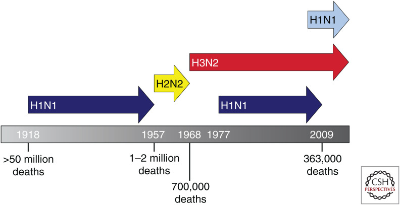
Just over a century ago in 1918-1919, the "Spanish" influenza pandemic appeared nearly simultaneously around the world and caused extraordinary mortality-estimated at 50-100 million fatalities-associated with unexpected clinical and epidemiological features. The pandemic's sudden appearance and high fatality rate were unprecedented, and 100 years later still serve as a stark reminder of the continual threat influenza poses. Sequencing and reconstruction of the 1918 virus have allowed scientists to answer many questions about its origin and pathogenicity, although many questions remain. Several of the unusual features of the 1918-1919 pandemic, including age-specific mortality patterns and the high frequency of severe pneumonias, are still not fully understood. The 1918 pandemic virus initiated a pandemic era still ongoing. The descendants of the 1918 virus remain today as annually circulating and evolving influenza viruses causing significant mortality each year. This review summarizes key findings and unanswered questions about this deadliest of human events.


Figures

Figure 1.
Figure 1.
Influenza pandemics of the past century. The 1918 “Spanish flu” pandemic was caused by a founder H1N1 influenza A virus. The three subsequent pandemics of 1957, 1968, and 2009 resulted from descendants of the 1918 virus, which acquired one or more genes through reassortment (Morens et al. 2009). Arrows reflect the years of annual epidemics of seasonal influenza circulation that occurred after each pandemic. In 1977, pre-1957 human H1N1 viruses reemerged presumably through accidental release of an older human strain (Nakajima et al. 1978). This resulted in a 20-year gap in H1N1 circulation as indicated by the discontinuous blue line. Consequently, human H3N2 viruses cocirculated with human 1918-lineage seasonal H1N1 viruses from 1977 until 2009, when this lineage was replaced by the novel swine-origin H1N1 2009 pandemic virus. Since 2009, H3N2 viruses have cocirculated with 2009 pandemic-lineage H1N1 viruses in humans.
Figure 2.
Figure 2.
Pathologic features of the 1918 virus. (A) 1918 influenza viral antigens were observed throughout the entire respiratory tract in autopsy tissues of 1918 victims, including epithelial cells in the tracheobronchial tree (the brown stain on a light blue background shows an immunohistochemical stain for influenza viral antigens). (B) Influenza viral antigens were similarly seen in the alveolar lining cells and alveolar macrophages of the lung. (C) 1918 HA sequences from different human autopsy specimens contained polymorphisms that affect receptor-binding specificity. No differences were observed, however, in the distribution of viral antigen in autopsy tissues. This is supported by in vitro modeling to assess binding to human bronchial airway epithelial cells as imaged by confocal microscopy. The pseudocolors are influenza viral antigen (green), goblet cells (magenta), ciliated cells (red), basal cells (blue), and nuclei (gray). Scale bar, 20 µm. (D) Section of lung showing diffuse alveolar damage (DAD) with hyaline membranes lining alveoli. The alveolar airspaces contain edema fluid, strands of fibrin, desquamated epithelial cells, and inflammatory cells. Original magnification, 400×. (E) Section of lung with acute pneumonia, showing a bacterial bronchopneumonia pattern consisting of necrotizing bronchitis and bronchiolitis, and massive infiltration of neutrophils into the airspaces of surrounding alveoli. Original magnification, 100×. (F) Tissue Gram stain of a fatal 1918 influenza pneumonia case. Gram-positive cocci morphologically compatible with Streptococcus pneumoniae. Original magnification, 1000×. A, B, and DF represent histologic findings from 1918 autopsy cases (Reid et al. 2003). C shows a confocal image produced using methods as described in Walters et al. (2016).
Figure 3.
Figure 3.
Age-specific influenza mortality (1918–1922). The 1918 Spanish flu pandemic appeared in Breslau (now Wrocław, Poland) in October 1918, causing high mortality. The “W-shaped” age-specific mortality pattern shown here was seen worldwide. Influenza age-specific mortality is usually “U-shaped” with higher mortality in infants and the elderly. A third peak of mortality in young adults (peaking at about age 27) was uniquely associated with the 1918 pandemic. Pandemic recurrences 15 and 26 months later were, however, associated with lower overall mortality and the virtual disappearance by 1922 of mortality in young adults (created from data in Lubinski 1923).

References

    1. Andreasen V, Viboud C, Simonsen L. 2008. Epidemiologic characterization of the 1918 influenza pandemic summer wave in Copenhagen: implications for pandemic control strategies. J Infect Dis 197: 270–278. 10.1086/524065 - DOI - PMC - PubMed
    1. Barry JM. 2004. The site of origin of the 1918 influenza pandemic and its public health implications. J Transl Med 2: 3 10.1186/1479-5876-2-3 - DOI - PMC - PubMed
    1. Basler CF, Reid AH, Dybing JK, Janczewski TA, Fanning TG, Zheng H, Salvatore M, Perdue ML, Swayne DE, García-Sastre A, et al. 2001. Sequence of the 1918 pandemic influenza virus nonstructural gene (NS) segment and characterization of recombinant viruses bearing the 1918 NS genes. Proc Natl Acad Sci 98: 2746–2751. 10.1073/pnas.031575198 - DOI - PMC - PubMed
    1. Budd AP, Wentworth DE, Blanton L, Elal AIA, Alabi N, Barnes J, Brammer L, Burns E, Cummings CN, Davis T, et al. 2018. Update: influenza activity—United States, October 1, 2017–February 3, 2018. MMWR Morb Mortal Wkly Rep 67: 169–179. 10.15585/mmwr.mm6706a1 - DOI - PMC - PubMed
    1. Chandra S. 2013. Mortality from the influenza pandemic of 1918–19 in Indonesia. Popul Stud (Camb) 67: 185–193. 10.1080/00324728.2012.754486 - DOI - PMC - PubMed

MeSH terms

LinkOut - more resources