. 2020 Feb 5;40(6):1286-1301.
doi: 10.1523/JNEUROSCI.0961-19.2019. Epub 2019 Dec 23.

Neuronal Activity in the Primate Amygdala during Economic Choice

Affiliations

Neuronal Activity in the Primate Amygdala during Economic Choice

Ahmad Jezzini et al. J Neurosci. .

Abstract

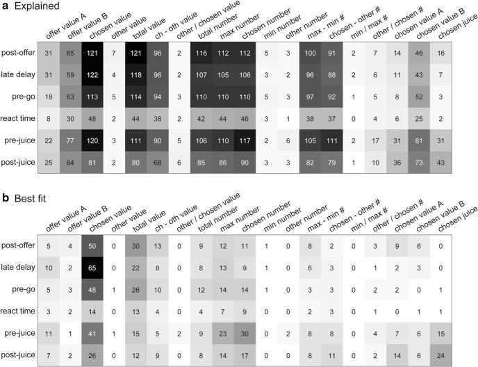
Multiple lines of evidence link economic choices to the orbitofrontal cortex (OFC), but other brain regions may contribute to the computation and comparison of economic values. A particularly strong candidate is the basolateral amygdala (BLA). Amygdala lesions impair performance in reinforcer devaluation tasks, suggesting that the BLA contributes to value computation. Furthermore, previous studies of the BLA have found neuronal activity consistent with a value representation. Here, we recorded from the BLA of two male rhesus macaques choosing between different juices. Offered quantities varied from trial to trial, and relative values were inferred from choices. Approximately one-third of BLA cells were task-related. Our analyses revealed the presence of three groups of neurons encoding variables offer value, chosen value, and chosen juice In this respect, the BLA appeared similar to the OFC. The two areas differed for the proportion of neurons in each group, as the fraction of chosen value cells was significantly higher in the BLA. Importantly, the activity of these neurons reflected the subjective nature of value. Firing rates in the BLA were sustained throughout the trial and maximal after juice delivery. In contrast, firing rates in the OFC were phasic and maximal shortly after offer presentation. Our results suggest that the BLA supports economic choice and reward expectation.SIGNIFICANCE STATEMENT Economic choices rely on the orbitofrontal cortex (OFC), but other brain regions may contribute to this behavior. A strong candidate is the basolateral amygdala (BLA). Previous results are consistent with a neuronal representation of value, but the role of the BLA in economic decisions remains unclear. Here, we recorded from monkeys choosing between juices. Neurons in the BLA encoded three decision variables: offer value, chosen value, and chosen juice These variables were also identified in the OFC. The two areas differed in the proportion of cells encoding each variable and in the activation timing. In the OFC, firing rates peaked shortly after offer presentation; in the BLA, firing rates were sustained and peaked after juice delivery. These results suggest that the BLA supports choices and reward expectation.

Keywords: decision making; neuroeconomics; neurophysiology; subjective value.


Figures

Figure 1.
Figure 1.
Experimental design and choice patterns. a, Task design. At the beginning of the trial, the animal fixated on a central location. Two offers represented by sets of colored squares appeared on the two sides of a fixation point. After a randomly variable delay (1–2 s), two saccade targets appeared by the offers (go signal). The animal indicated its choice with a saccade and maintained peripheral fixation for 0.75 s, after which the chosen juice was delivered. b, Time windows. c, Example choice pattern. The percentage of B choices (y-axis) is plotted against the log quantity ratio (log(qB/qA), x-axis). For this session, the probit regression indicated ρ = 2.12. d MRI scan from monkey H with the recording electrode placed in the basolateral amygdala. The asterisk indicates the tip of the electrode.
Figure 2.
Figure 2.
Reconstruction of recording sites. Locations are shown on a 1 mm grid. The x-axis and y-axis represent mediolateral and dorsoventral coordinates, respectively. Rostrocaudal locations were collapsed. Mediolateral coordinates are interaural estimates obtained by aligning the recording grid to the postsurgery MRI. Dorsoventral coordinates indicate the depth from the dura (as opposed to interaural coordinates). Dashed lines indicate our best estimate of the boundaries between different nuclei. B, L, and Ce indicate basal, lateral, and central nucleus, respectively. a, b, Recording locations for monkey H (a) and monkey I (b). For each location, the circle radius represents the number of recorded cells (see legend), and colors indicate the proportions of task-related versus untuned cells. c, d, Map of cell types for monkey H (c) and monkey I (d). For each location, the circle radius represents the number of task-related cells (see legend). Gray shades indicate the fractions of cells classified as offer value (gray), chosen value (blue), or chosen juice (yellow).
Figure 3.
Figure 3.
Example of neuronal responses. a, Response encoding the chosen value. Left, Black dots indicate the choice pattern and red symbols indicate firing rates. Diamonds and circles indicate trials in which the animal chose juice A and juice B, respectively. Right, The same neuronal response is plotted against the variable chosen value. The black line is from a linear regression (R2 is indicated). b, Response encoding the chosen value (negative encoding). In this case, the firing rate of the cell decreased as a function of the chosen value. c, Response encoding offer value A. d, Response encoding offer value B. e, Response encoding the chosen juice. bd, Conventions are as in a. ae, Responses are from the following time windows: postoffer (a), prego (b), postjuice (c), postoffer (d), postjuice (e).
Figure 4.
Figure 4.
Population results of the linear regression analysis. a, Explained responses. Each number indicates the number of responses explained by one variable in one time window. For example, in the postoffer time window, the variable chosen value explained 121 responses. Of note, because variable chosen value is highly correlated with other variables (total value, chosen number, etc.), many of these responses contributed to multiple bins in this plot. Different shades of gray represent the same results indicated numerically. b, Best fit. Here, numbers indicate the number of responses for which the corresponding variable provided the best fit (highest R2 value). In this panel, each response appears in one bin at most. Here, the variable chosen value clearly dominates across time windows.
Figure 5.
Figure 5.
Variable selection analysis. a, Stepwise procedure. Top, Same as in Figure 4b (using the same gray scale; see legend of Fig. 4b). In the first three iterations, the procedure selects the variables chosen value, chosen juice, and offer value A|B (asterisk indicates selected variables). Variables selected in subsequent iterations did not reach the 2% criterion (dot indicates selected-and-discarded variables). b, Percentage of explained responses, stepwise procedure. The panel illustrates the percentage of responses explained as a function of the number of selected variables. Here, 100% on the y-axis represents the total number of task-related responses (N = 863). The dotted line indicates the number of responses explained by the 19 variables (N = 804). Collectively, the three variables offer value A|B, chosen value, and chosen juice explained 759 responses. c, Total R2 value explained, best-subset procedure. The total R2 value (y-axis) is expressed as a percentage of the theoretical maximum, defined as sum(R2max) across the population. For each cell, R2max is the highest R2 value provided by one of the 19 variables. d, Distribution of R2 values. Each response was classified as encoding one of the variables offer value A|B, chosen value, and chosen juice. Notably, many more responses encoded the chosen value compared with other variables. Each histogram represents the distribution of R2 values for the corresponding variable.
Figure 6.
Figure 6.
Categorical encoding. For each pair of selected variables, we examined the difference in R2 values. a, Variables offer value and chosen value. We identified responses encoding offer value A or chosen value. Because many more responses encoded the latter, we randomly down sampled so that the two populations were of equal numbers. For each response, we computed R2offer value A and R2chosen value. We then repeated these operations for responses encoding the offer value B or the chosen value, and we pooled the results obtained for the two juices A and B. We then plotted R2offer value against R2chosen value (left) and the distribution for the difference ΔR2 (right). The distribution for ΔR2 presented a significant dip (p = 0, Hartigan's dip test), indicating that neuronal responses encoding variables offer value and chosen value were categorically distinct. b, Variables chosen value and chosen juice. The distribution for ΔR2 presented a significant dip (p = 0, Hartigan's dip test). c, Variables offer value and chosen juice. Same procedures as for a. The dip in the distribution for ΔR2 did not reach the significance threshold (p = 0.2, Hartigan's dip test). We repeated these analyses separately for each monkey and obtained very consistent results. For the three pairs of variables, Hartigan's dip test indicated p = 0.001, p = 0.001 and p = 0.2 for monkey H, and p = 0.024, p = 0.001 and p = 0.29 for monkey I.
Figure 7.
Figure 7.
U-shaped responses reflect the subjective nature of value. a, Example response. Left, Conventions are as in Figure 3a. Right, Firing rates were regressed on the chosen juice quantity, separately for trial types in which the animal chose juice A and juice B. We thus obtained the two slopes βA and βB. The slope ratio provided a neuronal measure for the relative value of the juices ρneuronal = βAB, which we compared with the behavioral measure obtained from the probit regression. For this response, ρbehavioral = 2.2 and ρneuronal = 2.3 ± 1.0 (mean ± SEM; SEM calculated through error propagation). b, Population analysis. Here, ρneuronal and ρbehavioral are plotted against each other in log-log scale. Each symbol represents one response, different colors indicate different juice pairs (see legend), and regression lines show the results of the ANCOVA (full model). In the legend, H and I identify the animal. In total, 427 responses from 16 juice pairs were included in the analysis. c, Estimates for individual juice pairs. Left, right, The estimate obtained from the ANCOVA for the intercept (left) and for the slope (right), separately for each juice pair. The association between juice pairs and color symbols is defined in b. Values on the x-axes refer to the relation between log(ρneuronal) and log(ρbehavioral). Vertical dotted lines (intercept = 0, slope = 1) correspond to the values predicted by the identity ρneuronal = ρbehavioral. Error bars indicate 3 SEs, and empty circles highlight juice pairs for which, by this measure, data points differ significantly from the values 0 (intercept) and 1 (slope). For 13 of 16 pairs, the relation between ρneuronal and ρbehavioral was indistinguishable from identity. Note that very similar results were obtained for the two animals.
Figure 8.
Figure 8.
Population activity profiles in the BLA. a, b, Activity profiles for the population of chosen value cells with positive encoding (a) and negative encoding (b). To calculate activity profiles, trials were separately aligned at offer and juice delivery. Trials were divided into three groups according to the chosen value. For each trial, the spike train was smoothed using a kernel that mimicked the postsynaptic potential by exerting influence only forward in time (decay time constant, 20 ms). Resulting traces were averaged across trials and across cells. The three traces refer to low, medium, and high chosen values. c, Activity profile for the population of offer value cells with positive encoding. The three traces refer to low, medium, and high values of the encoded juice. d. Activity profile of the population of chosen juice cells. The two traces correspond to trials in which the animal chose the encoded juice or the other juice.
Figure 9.
Figure 9.
Population activity profiles in the OFC. Same format and conventions as in Figure 8. ad Activity profiles for the populations of chosen value cells with positive encoding (a), chosen value cells with negative encoding (b), offer value cells with positive encoding (c) and chosen juice cells (d).
Figure 10.
Figure 10.
Distribution of baseline activity in the amygdala and OFC. Histograms show the distributions of mean firing rates recorded during the 500 ms preceding the offer (preoffer time window) in the BLA (yellow) and OFC (blue). Note that firing rates are plotted on a log scale. Green color indicates overlap between the two distributions. Across the population, the baseline activity recorded in the amygdala was significantly higher than that recorded in the OFC (p < 10−20, Kruskal–Wallis test). This result held true in each animal (both p < 10−20).
Figure 11.
Figure 11.
Neuronal encoding of decision variables, temporal profile. a, Amygdala, offer value cells (N = 55). Each cell was examined in 250 ms sliding time windows shifted by 50 ms. Time windows were separately aligned with offer onset (left) and juice delivery (right). In each window, a linear regression of firing rates against the offer value provided a p value for the regression slope. The smaller the p value was, the stronger the encoding was. For each cell and for each time window, we computed LP = −log10(p). The panel shows, for each cell, LP over time. To interpret gray shades, see the legend on the right. Note that LP = 0, 2, and 4 correspond to p = 1, p = 0.01, and p = 10−4, respectively. In the plot, cells were ranked from bottom to top according to the tuning latency. b, Amygdala, chosen value cells (N = 214). c, OFC, offer value cells (N = 61). d, OFC, chosen value cells (N = 66). bd, All conventions are as in a.
Figure 12.
Figure 12.
Tuning latencies in the amygdala and OFC. a, Offer value cells. b, Chosen value cells. In each panel, the results for the BLA and OFC are illustrated in red and blue, respectively.

References

    1. Amaral DG, Price JL (1984) Amygdalo-cortical projections in the monkey (Macaca fascicularis). J Comp Neurol 230:465–496. 10.1002/cne.902300402 - DOI - PubMed
    1. Amemori K, Graybiel AM (2012) Localized microstimulation of primate pregenual cingulate cortex induces negative decision-making. Nat Neurosci 15:776–785. 10.1038/nn.3088 - DOI - PMC - PubMed
    1. Balleine BW, Killcross AS, Dickinson A (2003) The effect of lesions of the basolateral amygdala on instrumental conditioning. J Neurosci 23:666–675. 10.1523/JNEUROSCI.23-02-00666.2003 - DOI - PMC - PubMed
    1. Belova MA, Paton JJ, Salzman CD (2008) Moment-to-moment tracking of state value in the amygdala. J Neurosci 28:10023–10030. 10.1523/JNEUROSCI.1400-08.2008 - DOI - PMC - PubMed
    1. Bermudez MA, Schultz W (2010) Reward magnitude coding in primate amygdala neurons. J Neurophysiol 104:3424–3432. 10.1152/jn.00540.2010 - DOI - PMC - PubMed

Publication types

LinkOut - more resources