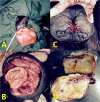
Surgical management of penile sarcoid in a stallion
- PMID: 31871412
- PMCID: PMC6920054
- DOI: 10.1294/jes.30.99
Surgical management of penile sarcoid in a stallion
Abstract
This report describes surgical management and breeding implications of a case of penile sarcoid associated with penis laceration in a 4-year-old Kurdish stallion. A large fleshy mass on the distal end of the penis that resulted in urethral meatus deviation and dysuria was detected in a physical examination. No evidence of local extent or metastasis was detected. Under general anaesthesia, the involved distal portion of the penis was removed through partial phallectomy. Histopathological examination of the mass confirmed a fibroblastic sarcoid. Partial phallectomy was successful for management of penile sarcoid and resulted in no postoperative complications or tumour recurrence in long-term follow up; however, successful ejaculation and semen collection have not been achieved.
Keywords: partial phalectomy; penis; sarcoid; stallion.
©2019 The Japanese Society of Equine Science.
Figures


References
-
- Brinsko S.P. 1998. Neoplasia of the male reproductive tract. Vet. Clin. North Am. Equine Pract. 14: 517–533. - PubMed
-
- Brinsko S.P. 2011. Semen collection techniques and insemination procedures. pp. 1268–1277. In: Equine Reproduction, 2nd ed. (McKinnon, A.O., and Squires, E.L. eds.), John Wiley & Sons, Ames.
-
- Byrne D.P., Woolford L., Booth T.M. 2014. Penile haemangiosarcoma in a breeding stallion. Equine Vet. Educ. 28: 304–309.
-
- Crump J., Jr, Crump J. 1989. Stallion ejaculation induced by manual stimulation of the penis. Theriogenology 31: 341–346. - PubMed
-
- Davies Morel M.C.G. 1999. Equine Artificial Insemination. pp. 151–189. CABI Pub., New York.
LinkOut - more resources
Full Text Sources
