Review
. 2019 Oct 23;6(24):1901779.
doi: 10.1002/advs.201901779. eCollection 2019 Dec.

Extracellular Vesicles in Cancer Immune Microenvironment and Cancer Immunotherapy

Affiliations
Review

Extracellular Vesicles in Cancer Immune Microenvironment and Cancer Immunotherapy

Feng Xie et al. Adv Sci (Weinh). .

Abstract

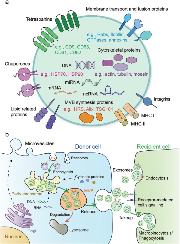
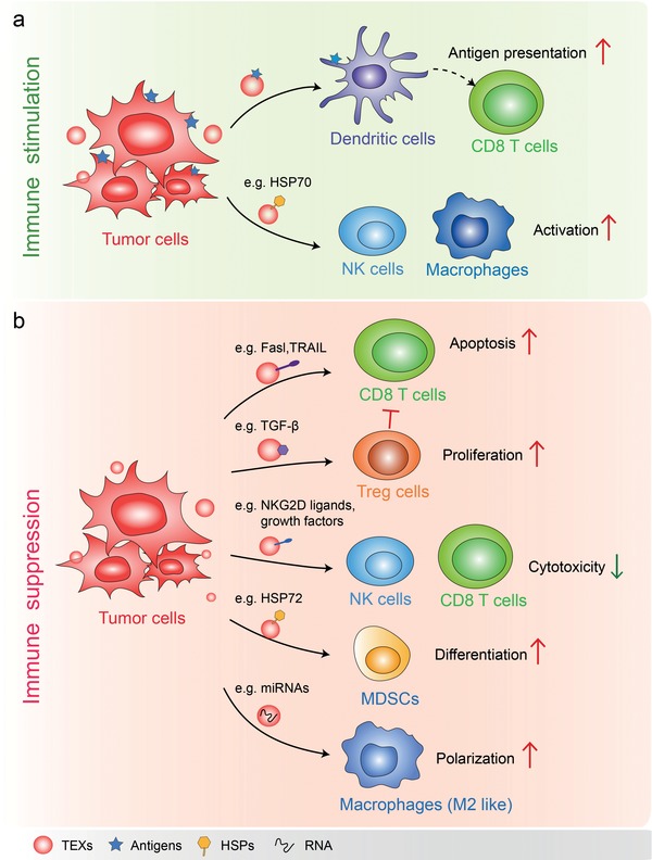
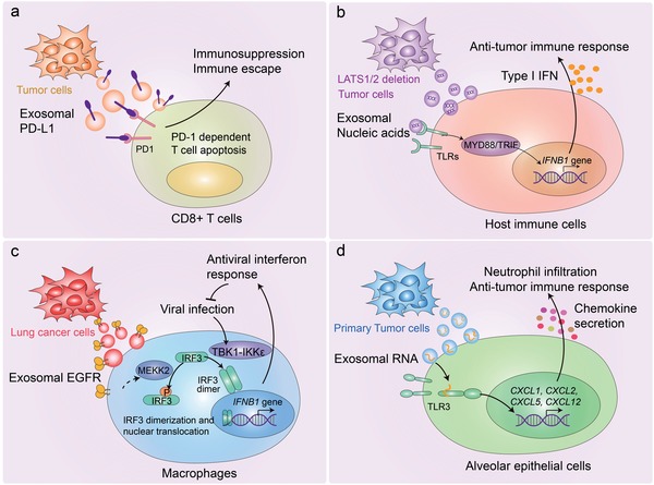
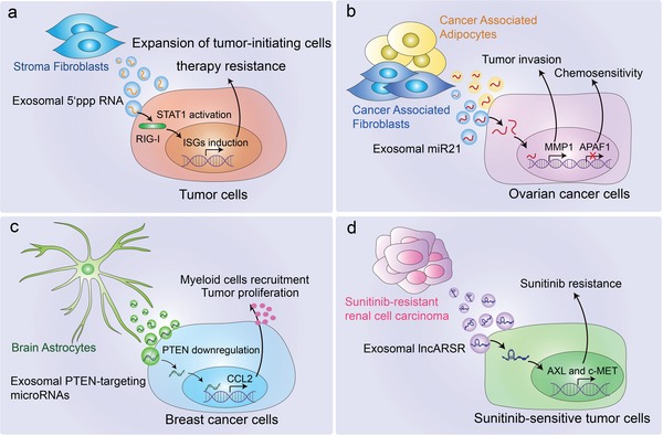
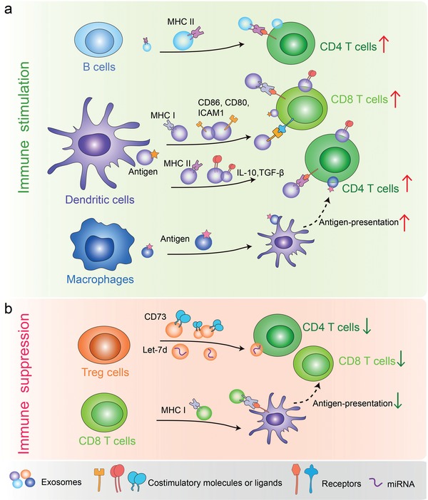
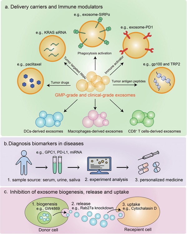
Extracellular vesicles (EVs) are secreted by almost all cells. They contain proteins, lipids, and nucleic acids which are delivered from the parent cells to the recipient cells. Thereby, they function as mediators of intercellular communication and molecular transfer. Recent evidences suggest that exosomes, a small subset of EVs, are involved in numerous physiological and pathological processes and play essential roles in remodeling the tumor immune microenvironment even before the occurrence and metastasis of cancer. Exosomes derived from tumor cells and host cells mediate their mutual regulation locally or remotely, thereby determining the responsiveness of cancer therapies. As such, tumor-derived circulating exosomes are considered as noninvasive biomarkers for early detection and diagnosis of tumor. Exosome-based therapies are also emerging as cutting-edge and promising strategies that could be applied to suppress tumor progression or enhance anti-tumor immunity. Herein, the current understanding of exosomes and their key roles in modulating immune responses, as well as their potential therapeutic applications are outlined. The limitations of current studies are also presented and directions for future research are described.

Keywords: anti‐tumor immunity; cancer immunotherapy; exosomes; extracellular vesicles; tumor microenvironment.


Conflict of interest statement

The authors declare no conflict of interest.

Figures

Figure 1
Figure 1
Molecular composition, biogenesis, secretion, and uptake of the exosomes. a) Exosomes contain complex contents including proteins, mRNA, miRNA, ncRNA, and DNA. TSG101 and Alix are involved in the formation of internal vesicles of MVBs. The tetraspanins such as CD9, CD63, and CD81, are the markers currently used to characterize exosomes. b) Exosomes originate from ILVs in MVBs. Firstly, proteins are transported from the Golgi or internalized from the cell surface, and nucleic acids should be endocytosed and transferred into the early endosomes. Then early endosomes maturate into late endosomes/MVB, which follow either the secretory or the degradative pathway. Microvesicles are released after formation by budding from the cytomembrane. Once released, exosomes can interact with recipient cells by direct signaling through ligand/receptor molecules on their respective surfaces. Exosomes can also be taken up by recipient cells via different manners such as direct fusion of their membrane, endocytosis, macropinocytosis, and even phagocytosis (right). Thus, exosomes function as a mode of intercellular communication and molecular transfer.
Figure 2
Figure 2
Functions of TEXs in tumor immune environment. a) TEXs present tumor antigen and enhance anti‐tumor immunity: in the presence of dendritic cells, TEXs loaded with specific antigens are capable of promoting the activation of tumor antigen‐specific CD8+ cytotoxic T‐lymphocytes. The HSP70 surface‐positive TEXs stimulate migratory and cytolytic activity of NK cells and macrophages. b) In most cases, TEXs function as immune suppressor. For instance, TEXs containing Fasl or TRAIL induce the apoptosis of T cells and suppress activation of T cells. TEXs bearing TGF‐β increase the proliferation of Treg cells which suppress immune responses. TEXs expressing NKG2D ligands or TGF‐β1 can inhibit the cytotoxicity of NK cells and CD8 T cells by triggering down‐regulation of their surface NKG2D expression. HSP72 bearing TEXs trigger STAT3 activation in MDSCs and promote MDSCs suppressive functions. TEXs containing miRNAs such as miR‐21‐3p, miR‐125b‐5p, miR‐181d‐5p, and miR‐1246 remodel macrophages to a tumor‐promoted phenotype.
Figure 3
Figure 3
Mechanisms of TEXs in modulating innate and adaptive immunity. a) Tumor cell‐derived exosomal PD‐L1 can be transferred to CD8+ T cells, leading to the immunosuppression and immune escape in melanoma and prostate cancer. b) LATS1/2 deficient tumor cells secrete nucleic‐acid‐rich extracellular vesicles, which induces anti‐tumor immune responses via type I interferon. c) Tumor cell‐derived exosomal EGFR can be transferred into host macrophages to reduce their production of type I interferon and inhibit antiviral immunity. d) Primary tumor‐derived exosomal small nuclear RNAs can be transferred to the lung epithelial cells, leading to the activation of TLR3, production of chemokine, and recruitment of neutrophils. Thus, tumor‐derived exosomal small nuclear RNAs can elicit a pro‐metastatic inflammatory microenvironment by suppressing innate and adaptive anti‐tumor immunity.
Figure 4
Figure 4
Stroma cells in the TME support tumor progression via secreting exosomes. a) NOTCH‐MYC signaling in stromal fibroblasts shed exosomes containing unshielded RN7SL1 RNA. Upon being transferred to breast cancer cells, unshielded RN7SL1 activates RIG‐I and STAT1, and increases ISG induction, resulting in tumor growth, metastasis, and therapy resistance. Upon being transferred to immune cells, it can also drive an inflammatory response by increasing the percentage of myeloid DC populations. b) Cancer‐associated adipocytes and fibroblast‐derived exosomal miR21 can be transferred to the cancer cells, which downregulate APAF1 expression and upregulate MMP1 expression, resulting in tumor invasion and chemoresistance. c) Brain astrocyte‐derived exosomal PTEN‐targeting microRNAs can be transferred to metastatic tumor cells, induce an increased secretion of the chemokine CCL2 and facilitate the recruitment of IBA1+ myeloid cells which promotes tumor outgrowth. d) Sunitinib resistant RCC cell‐derived exosomal lncARSR can be transferred to sensitive cells and facilitates AXL and c‐MET expression, thus disseminating sunitinib resistance.
Figure 5
Figure 5
Exosomes from distinct immune cells play divergent roles in regulating cancer immunity. a) B cell‐derived exosomes bearing MHC II activate CD4+ T cells. DC‐derived exosomes containing tumor‐derived antigens, costimulatory molecules, and proteins, can promote the activation of CD4+ T cells and CD8+ T cells. Macrophage‐derived exosomes bearing MHC I can be transferred to DCs, thereby enabling them to activate antigen‐specific CD4+ T cells. b) Treg‐derived exosomes containing CD73 can inhibit T cell activation. CD8+ T cell‐derived exosomes carrying MHC I also mediate immune suppression by inhibiting the antigen presentation of DCs.
Figure 6
Figure 6
Exosomes in cancer immunotherapy. a) As exosomes have high stability in circulation and good capacity to transfer horizontal cargo, they have been explored as delivery carriers loaded with drugs or tumor targeted RNAi in different diseases. In addition, exosomes can be employed as immune modulators by expressing proteins such as SIRPα, PD1, or tumor antigen peptides. The exosomes derived from immune cells including DCs, macrophages and CD8+ T cells are demonstrated to stimulate anti‐tumor immune responses. Importantly, large‐scale generation of good manufacturing practice‐grade (GMP‐grade) and clinical‐grade exosomes are generated for clinical applications. b) Exosomes bearing GPC1, PD‐L1, or certain miRNA could be valuable as cancer biomarkers. c) Inhibition of exosomes biogenesis, release, and uptake is another strategy of cancer immunotherapy.

References

    1. Trams E. G., Lauter C. J., Salem N. Jr., Heine U., Biochim. Biophys. Acta, Biomembr. 1981, 645, 63. - PubMed
    1. Simons M., Raposo G., Curr. Opin. Cell Biol. 2009, 21, 575. - PubMed
    1. Thery C., Ostrowski M., Segura E., Nat. Rev. Immunol. 2009, 9, 581. - PubMed
    1. Zabeo D., Cvjetkovic A., Lasser C., Schorb M., Lotvall J., Hoog J. L., J. Extracell. Vesicles 2017, 6, 1329476. - PMC - PubMed
    1. Zhang H., Freitas D., Kim H. S., Fabijanic K., Li Z., Chen H., Mark M. T., Molina H., Martin A. B., Bojmar L., Fang J., Rampersaud S., Hoshino A., Matei I., Kenific C. M., Nakajima M., Mutvei A. P., Sansone P., Buehring W., Wang H., Jimenez J. P., Cohen‐Gould L., Paknejad N., Brendel M., Manova‐Todorova K., Magalhaes A., Ferreira J. A., Osorio H., Silva A. M., Massey A., Cubillos‐Ruiz J. R., Galletti G., Giannakakou P., Cuervo A. M., Blenis J., Schwartz R., Brady M. S., Peinado H., Bromberg J., Matsui H., Reis C. A., Lyden D., Nat. Cell Biol. 2018, 20, 332. - PMC - PubMed

LinkOut - more resources