

Adult primary gastric volvulus, a report of two cases
- PMID: 31872178
- PMCID: PMC6917551
- DOI: 10.21037/acr.2019.10.03
Adult primary gastric volvulus, a report of two cases
Abstract
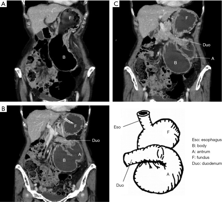
Gastric volvulus is the medical situation that a stomach is twisted beyond the physiological range. It is a rare disease which is hard to experience in routine medical examination. Principally surgical treatment is essential for the acute type. However, the conservative therapy should be attempted in some cases, such as decompression of a stomach with a nasogastric tube, endoscopic reduction and so forth. Concerning surgical operation, the base is reduction of the torsion and immobilization of stomach. Recently, laparoscopic surgery is performed for the case that the general condition is stable or chronically progressive in the early stages. Percutaneous endoscopic gastrostomy (PEG) had also been performed for gastric immobilization. However, the recurrences and problems of twisting around the gastrostomy site were reported in addition to the problem of cosmetic outcomes. Therefore, the case is decreasing. In this paper, we present two cases on adult primary gastric volvulus. For the first case, endoscopic reduction was not good enough to release the torsion state. Then laparoscopic gastropexy was performed successfully. For the second case, we succeeded in endoscopic reduction. Since the patient had already experienced gastric volvulus, laparoscopic surgery was performed. The upper and middle gastric bodies were secured to the anterior abdominal wall, and gastric antrum to the ligamentum teres hepatis with interrupted absorbable sutures respectively. However, partial gastric volvulus recurred after ten and a several days postoperatively due to cutting off of the suture at the antrum secured to the ligamentum teres hepatis at previous surgery. Then, PEG for 2 points of lower body and antrum were performed to secure the antrum. The gastrostomies were removed 6 months after the surgery. Immobilization by laparoscopic gastropexy and PEG are useful for gastric volvulus due to their significant merit of minimum invasiveness. Concerning gastropexy, the number of sutures is very important for the secured part not to be torn off.
Keywords: Gastric volvulus; gastropexy; laparoscopic surgery; percutaneous endoscopic gastrostomy (PEG).
2019 AME Case Reports. All rights reserved.
Conflict of interest statement
Conflicts of Interest: The authors have no conflicts of interest to declare.
Figures


Similar articles
-
A Case of Laparoscopic-Assisted Percutaneous Endoscopic Gastrostomy (LAPEG) for Gastric Volvulus.Case Rep Med. 2019 Dec 3;2019:3468084. doi: 10.1155/2019/3468084. eCollection 2019. Case Rep Med. 2019. PMID: 31871462 Free PMC article.
-
A combined laparoscopic and endoscopic approach to acute primary gastric volvulus.J Laparoendosc Adv Surg Tech A. 1997 Jun;7(3):177-81. doi: 10.1089/lap.1997.7.177. J Laparoendosc Adv Surg Tech A. 1997. PMID: 9448130
-
Laparoscopic suture gastropexy for gastric volvulus: a report of 14 cases.Surg Endosc. 2007 Jun;21(6):863-6. doi: 10.1007/s00464-006-9089-4. Epub 2006 Dec 16. Surg Endosc. 2007. PMID: 17180266
-
[Acute gastric volvulus: late complication of Nissen fundoplication. Report of two cases and review of the literature].Cir Cir. 2014 Sep-Oct;82(5):541-50. Cir Cir. 2014. PMID: 25259434 Review. Spanish.
-
Emergent laparoscopic reduction of acute gastric volvulus with anterior gastropexy.Surg Laparosc Endosc Percutan Tech. 2003 Dec;13(6):389-91. doi: 10.1097/00129689-200312000-00009. Surg Laparosc Endosc Percutan Tech. 2003. PMID: 14712102 Review.
Cited by
-
Gastric volvulus with necrosis and gangrene associated with wandering spleen: A rare case report from Syria.SAGE Open Med Case Rep. 2024 Jun 14;12:2050313X241262141. doi: 10.1177/2050313X241262141. eCollection 2024. SAGE Open Med Case Rep. 2024. PMID: 38881967 Free PMC article.
References
Publication types
LinkOut - more resources
Full Text Sources