. 2020 Jan 24;5(1):34-39.
doi: 10.1021/acssensors.9b00796. Epub 2020 Jan 8.

Enhanced Molecular Tension Sensor Based on Bioluminescence Resonance Energy Transfer (BRET)

Affiliations

Enhanced Molecular Tension Sensor Based on Bioluminescence Resonance Energy Transfer (BRET)

Eric J Aird et al. ACS Sens. .

Abstract

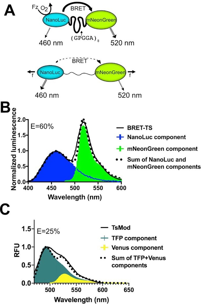
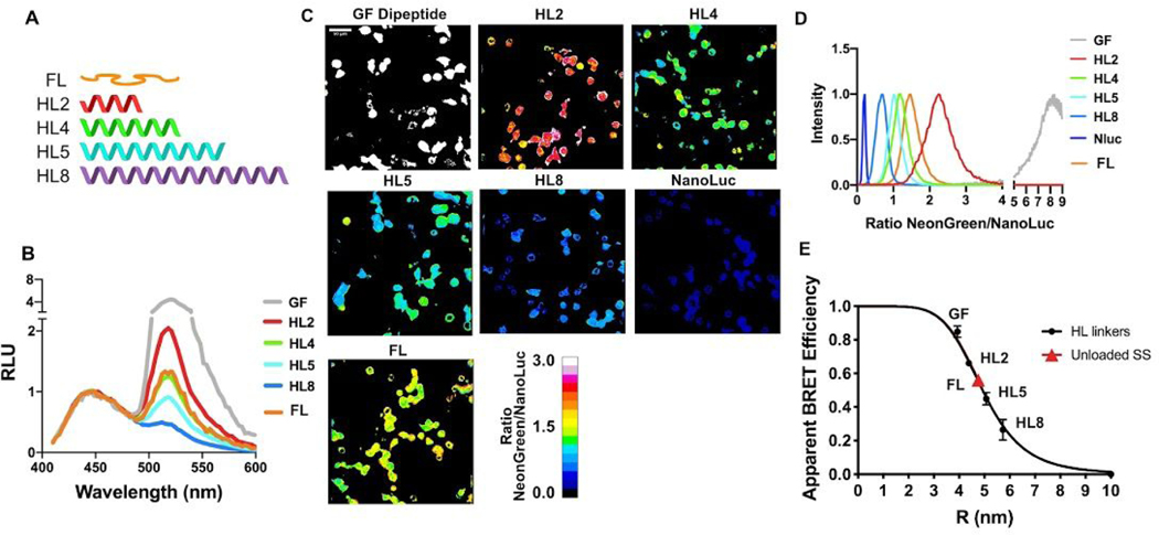
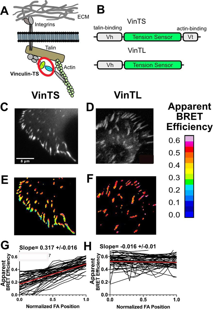
Molecular tension sensors measure piconewton forces experienced by individual proteins in the context of the cellular microenvironment. Current genetically encoded tension sensors use FRET to report on extension of a deformable peptide encoded in a cellular protein of interest. Here, we present the development and characterization of a new type of molecular tension sensor based on bioluminescence resonance energy transfer (BRET), which exhibits more desirable spectral properties and an enhanced dynamic range compared to other molecular tension sensors. Moreover, it avoids many disadvantages of FRET measurements in cells, including autofluorescence, photobleaching, and corrections of direct acceptor excitation. We benchmark the sensor by inserting it into the canonical mechanosensing focal adhesion protein vinculin, observing highly resolved gradients of tensional changes across focal adhesions. We anticipate that the BRET tension sensor will expand the toolkit available to study mechanotransduction at a molecular level and allow potential extension to an in vivo context.

Keywords: BRET; NanoLuc; TSMod; bioluminescence; molecular tension sensor.


Figures

Figure 1.
Figure 1.
Schematic of the genetically-encodable BRET molecular tension sensor (BRET-TS). (A) NanoLuc, the energy donor, is excited via addition of furimazine (Fz) in the presence of oxygen. In the absence of force (f), resonance energy transfer occurs to the fluorescent acceptor, mNeonGreen. With applied force, the donor-acceptor pair is separated, reducing resonance energy transfer. Unloaded spectral and resonance energy transfer properties of (B) BRET-TS in comparison to (C) FRET-based TSMod. Spectral components and resulting best-fit additive spectrum depicted. Axes in units of luminescence normalized at 460 nm and relative fluorescence units (RFU).
Figure 2.
Figure 2.
Characterization of distance dependence of BRET-TS in vitro and in cells. (A) Rigid alpha helical linkers (HL) of varying lengths were inserted between the mNeonGreen-NanoLuc pair in addition to a minimal dipeptide GF linker. NanoLuc alone was also measured. (B) Spectral plate reader emissions of recombinantly expressed proteins, normalized to NanoLuc peak emission (460 nm). (C) HEK293T cells transfected with the various HL linker constructs inserted into a plasma membrane localized protein. Cells were imaged using NanoLuc and mNeonGreen specific spectral filters and ratiometric mNeonGreen:NanoLuc images are shown. (D) Histograms of a field of cells from C were calculated and normalized to 1 to derive the average BRET ratio for a given linker. (E) Apparent RET vs R curves comparing HL linker BRET efficiency derived from in vitro recombinant protein BRET-TS and cell-surface BRET-TS versus distance r. R was calculated from apparent BRET efficiencies corrected for the ratio of acceptor to donor quantum yields (see Methods) and using the measured R0 least squares fit to 1/R6 function.
Figure 3.
Figure 3.
Measuring tension across vinculin. (A) Schematic of vinculin with inserted BRET tension sensor (BRET-TS) in the context of a focal adhesion. (B) Vinculin constructs used in these studies: full length vinculin with inserted BRET-TS (VinTS) and actin binding deficient vinculin with BRET-TS (VinTL). (C–D) Unprocessed luminescent images of focal adhesions. (E–F) Processed, ratiometric images from (C–D). Scale bar displayed as apparent BRET efficiency (out of 1 maximum). (G–H) Manual line scans taken across focal adhesions, normalized for focal adhesion (FA) length (n=30). The red line represents the linear regression of all line scans combined. The slope of the line is given +/− the standard error of the linear regression calculation. Images and line scans are representative from images collected over numerous independent experiments.

References

    1. Janmey PA; Miller RT Mechanisms of Mechanical Signaling in Development and Disease. J. Cell Sci 2011, 124 (1), 9–18. 10.1242/jcs.071001. - DOI - PMC - PubMed
    1. Bonnans C; Chou J; Werb Z. Remodelling the Extracellular Matrix in Development and Disease. Nat. Rev. Mol. Cell Biol 2014, 15 (12), 786–801. 10.1038/nrm3904. - DOI - PMC - PubMed
    1. Insua-Rodríguez J; Oskarsson T. The Extracellular Matrix in Breast Cancer. A dv. Drug Deliv. Rev 2016, 97, 41–55. 10.1016/j.addr.2015.12.017. - DOI - PubMed
    1. Wang N. Review of Cellular Mechanotransduction. J. Phys. D Appl. Phys 2017, 50 233002. 10.1088/1361-6463/aa6e18 - DOI - PMC - PubMed
    1. Humphrey JD; Dufresne ER; Schwartz MA Mechanotransduction and Extracellular Matrix Homeostasis. Nat. Rev. Mol. Cell Biol 2014, 15 (12), 802–812. 10.1038/nrm3896. - DOI - PMC - PubMed

Publication types

MeSH terms