. 2019 Dec 23;9(1):19643.
doi: 10.1038/s41598-019-55819-y.

Caveolin-1 gene therapy inhibits inflammasome activation to protect from bleomycin-induced pulmonary fibrosis

Affiliations

Caveolin-1 gene therapy inhibits inflammasome activation to protect from bleomycin-induced pulmonary fibrosis

Xin Lin et al. Sci Rep. .

Abstract

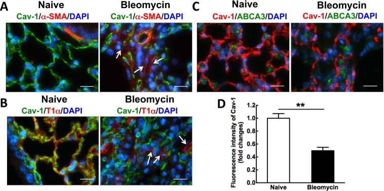
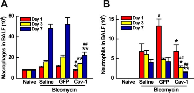
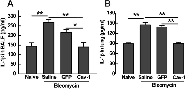
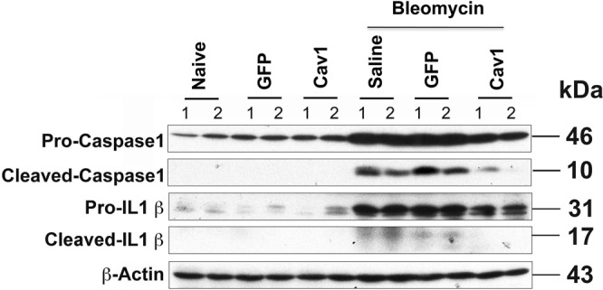
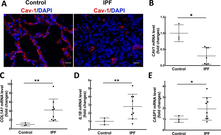
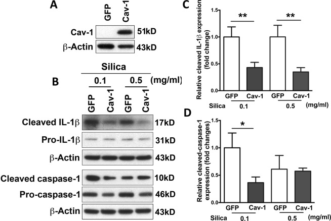
Idiopathic pulmonary fibrosis (IPF) is a devastating and fatal disease and characterized by increased deposition of extracellular matrix proteins and scar formation in the lung, resulting from alveolar epithelial damage and accumulation of inflammatory cells. Evidence suggests that Caveolin-1 (Cav-1), a major component of caveolae which regulates cell signaling and endocytosis, is a potential target to treat fibrotic diseases, although the mechanisms and responsible cell types are unclear. We show that Cav-1 expression was downregulated both in alveolar epithelial type I cells in bleomycin-injured mouse lungs and in lung sections from IPF patients. Increased expression of IL-1β and caspase-1 has been observed in IPF patients, indicating inflammasome activation associated with IPF. Gene transfer of a plasmid expressing Cav-1 using transthoracic electroporation reduced infiltration of neutrophils and monocytes/macrophages and protected from subsequent bleomycin-induced pulmonary fibrosis. Overexpression of Cav-1 suppressed bleomycin- or silica-induced activation of caspase-1 and maturation of pro-IL-1β to secrete cleaved IL-1β both in mouse lungs and in primary type I cells. These results demonstrate that gene transfer of Cav-1 downregulates inflammasome activity and protects from subsequent bleomycin-mediated pulmonary fibrosis. This indicates a pivotal regulation of Cav-1 in inflammasome activity and suggests a novel therapeutic strategy for patients with IPF.


Conflict of interest statement

Dr. Sime reports grants from NIH, during the conduct of the study; grants from NIH, grants and personal fees from UCB, personal fees from Boehringer Ingelheim, personal fees from Intermune/roche, personal fees from GSK, outside the submitted work. No other authors have any competing interests.

Figures

Figure 1
Figure 1
Cav-1 expression is decreased in bleomycin-induced pulmonary fibrosis, primarily, in alveolar epithelial type I cells after bleomycin administration for 21 days. (A) Immunofluorescence analysis of Cav-1 expression in lung tissue sections (Cav-1, green; nucleus, blue; α-SMA, red). (B) Immunofluorescence analysis of T1α, an epithelial type I cell marker, and Cav-1 expression in bleomycin-induced fibrosis tissue sections, compared to normal lungs (Cav-1, green; nucleus, blue; T1α, red). (C) Immunofluorescence analysis of ABCA3, an epithelial type II cell marker, and Cav-1 expression in bleomycin-induced fibrosis tissue sections, compared to normal lungs (Cav-1, red; nucleus, blue; ABCA3, green). Arrows indicate that Cav-1 expression is decreased in α-SMA-positive cells or epithelial type I cells. Scale bar: 10 μm. (D) Fluorescence intensity of Cav-1 was quantified using Image J software in the tissue sections (n = 8). Statistical analysis was by unpaired t test (mean ± SD; n = 4 mice per group), **P < 0.01.
Figure 2
Figure 2
Electroporation-mediated gene transfer of Cav-1 protects from bleomycin-induced pulmonary fibrosis. 100 μg of plasmid expressing GFP-tagged Cav-1 driven by the UbC promoter in 50 μl was delivered intratracheally to C57BL/6 mice and electroporated at 200 V/cm using 8 pulses of 10 msec duration. One day later, bleomycin (2 Unit/kg) was given to the lungs and the lungs were harvested at 21 days and subjected to (A) immunofluorescence staining to determine co-localization of GFP with Cav-1 in healthy mouse lungs or (B) bleomycin-injured lungs (GFP, green; nucleus, blue; Cav-1, red) (Scale bar: 10 μm), (C) Real-time PCR of left lobe, (D) H&E staining and (E) Masson trichrome staining of right lung tissue. One representative example out of four is shown. Scale bar: 100 μm. (F) Fibrosis score was evaluated by the Ashcroft scale in the tissue sections (n = 4). Statistical analysis was by one-way ANOVA (mean ± SEM; n = 4 mice per group), *P < 0.05 or **P < 0.01.
Figure 3
Figure 3
Gene transfer of Cav-1 reduces neutrophils and macrophages in BALF of bleomycin-challenged mice. (A) Alveolar macrophages and (B) neutrophils were quantified in BALF at days 1, 3, and 7 after bleomycin instillation. Cav-1 gene delivery prior to bleomycin decreased macrophage and neutrophil infiltration at all times compared to mice receiving control GFP plasmid and bleomycin. Statistical analysis was by one-way ANOVA (mean ± SEM; n = 5 mice per group). *P < 0.05, *P < 0.01 vs vector control; #P < 0.05, # # P < 0.01 vs bleomycin only.
Figure 4
Figure 4
Gene transfer of Cav-1 decreases IL-1β production in both BALF and lungs of bleomycin-challenged mice. IL-1β production in BALF (A) and lung (B) was analyzed at day 1 after bleomycin administration measured by ELISA. Statistical analysis was by one-way ANOVA (mean ± SEM; n = 5), *P < 0.05 or **P < 0.01.
Figure 5
Figure 5
Gene transfer of Cav-1 suppresses bleomycin-induced inflammasome activation in mouse lungs 21 days after bleomycin administration. 100 μg of plasmid in 50 μl was delivered intratracheally to C57BL/6 mice and electroporated at 200 V/cm using 8 pulses of 10 msec duration. One day later, bleomycin (2 Unit/kg) was given to the lungs and the lungs were harvested at 21 days and subjected to western blot. Cropped blots were used in this figure. Original full-length blots are presented in Supplementary Figure S1.
Figure 6
Figure 6
Inflammasome activation is involved in IPF patients with decreased Cav-1 expression. (A) Reduced Cav-1 expression was analyzed in lung sections by Immunofluorescence staining, compared to control (Cav-1, red; nucleus, blue). Representative photographs were taken at 400X magnification. Scale bar: 20 μm. mRNA levels of Cav-1 (B), collagen I (C), IL-1β (D) and caspase-1 (E) were detected by real-time PCR in lung tissue samples from IPF patients (n = 7–9) and control tissues (n = 3). Mean ± SD. Statistical analysis was analyzed by student t test. *P < 0.05 or **P < 0.01 compared to control.
Figure 7
Figure 7
Overexpression of Cav-1 abolished silica-induced inflammasome activation in rat primary alveolar epithelial type I cells. Alveolar epithelial type II cells were isolated from naïve rat and transfected with Cav-1 or vector control. Four days later, primed for 5 hours with 1 mg/ml LPS, and then stimulated for 24 hours with silica. (A) Overexpression of Cav-1 was measured by western blot. Cropped blots were used in this figure. Original full-length blots are presented in Supplementary Figure S2A. (B) The presence of mature IL-1β and cleaved caspase-1 was analyzed in media supernatants, and pro-IL-1β and pro-caspase-1 were detected in cell extracts by western blot. Cropped blots were used in this figure. Original full-length blots are presented in Supplementary Figure S2B. The densitometry of western blot shows cleaved IL-1β (C) and cleaved caspase-1 (D). Statistical analysis was analyzed by student t test. *P < 0.05 compared to GFP control.

References

    1. Wynn TA. Integrating mechanisms of pulmonary fibrosis. J Exp Med. 2011;208:1339–1350. doi: 10.1084/jem.20110551. - DOI - PMC - PubMed
    1. Gasse P, et al. IL-1R1/MyD88 signaling and the inflammasome are essential in pulmonary inflammation and fibrosis in mice. J Clin Invest. 2007;117:3786–3799. - PMC - PubMed
    1. Raghu G, Weycker D, Edelsberg J, Bradford WZ, Oster G. Incidence and prevalence of idiopathic pulmonary fibrosis. Am J Respir Crit Care Med. 2006;174:810–816. doi: 10.1164/rccm.200602-163OC. - DOI - PubMed
    1. Khalil N, O’Connor R. Idiopathic pulmonary fibrosis: current understanding of the pathogenesis and the status of treatment. CMAJ. 2004;171:153–160. doi: 10.1503/cmaj.1030055. - DOI - PMC - PubMed
    1. Hosseinian N, Cho Y, Lockey RF, Kolliputi N. The role of the NLRP3 inflammasome in pulmonary diseases. Ther Adv Respir Dis. 2015;9:188–197. doi: 10.1177/1753465815586335. - DOI - PubMed

Publication types