

miR-192-5p suppresses the progression of lung cancer bone metastasis by targeting TRIM44
- PMID: 31873114
- PMCID: PMC6928221
- DOI: 10.1038/s41598-019-56018-5
miR-192-5p suppresses the progression of lung cancer bone metastasis by targeting TRIM44
Abstract
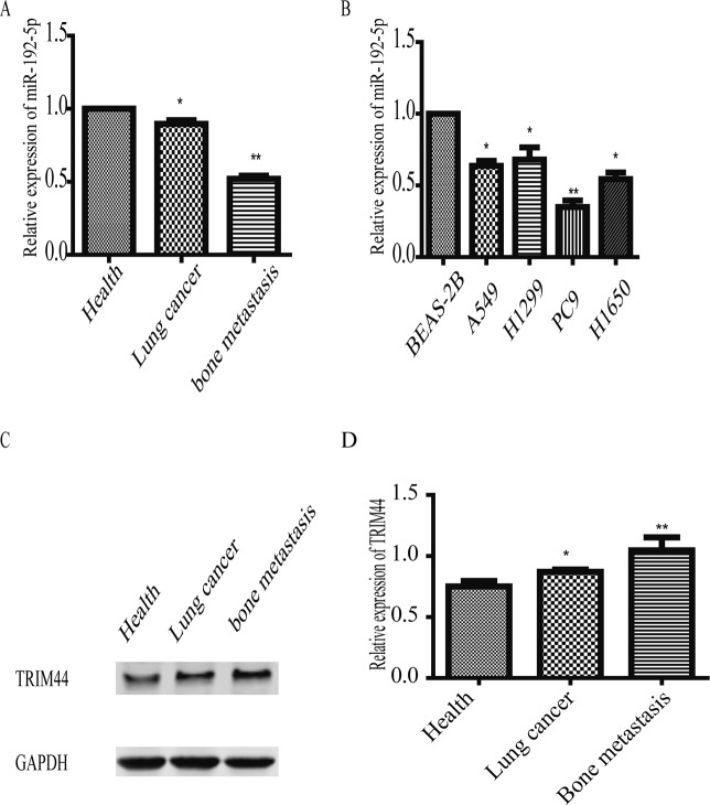
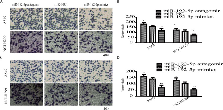
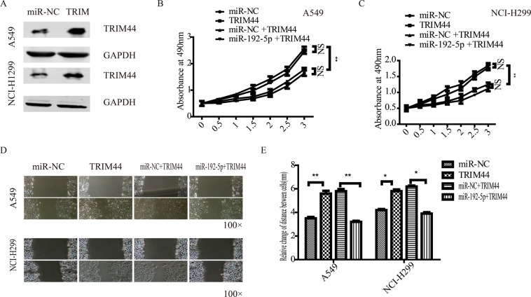
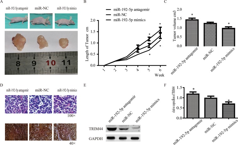
Lung cancer is the leading cause of cancer-related deaths worldwide, with 50-70% of patients suffering from bone metastasis. Accumulating evidence has demonstrated that miRNAs are involved in cell proliferation, migration, and invasion in malignancy, such as lung cancer bone metastasis. In the present study, we demonstrated that reduced miR-192-5p and increased TRIM44 levels were associated with the proliferation, migration and invasion of lung cancer. Furthermore, the potential functions of miR-192-5p were explored in A549 and NCI-H1299 cells. We found that miR-192-5p upregulation suppressed tumour behaviours in lung cancer cells. To further investigate whether miR-192-5p is associated with TRIM44, we used TargetScan software to predict the binding site between miR-192-5p and TRIM44. Luciferase activity assays were performed to verify this prediction. In addition, the significant role of miR-192-5p in negatively regulating TRIM44 expression was manifested by our research group. our results suggest that miR-192-5p inhibited the proliferation, migration and invasion of lung cancer through TRIM44.
Conflict of interest statement
The authors declare no competing interests.
Figures


Similar articles
-
Nm23-H1 inhibits lung cancer bone-specific metastasis by upregulating miR-660-5p targeted SMARCA5.Thorac Cancer. 2020 Mar;11(3):640-650. doi: 10.1111/1759-7714.13308. Epub 2020 Feb 5. Thorac Cancer. 2020. PMID: 32022430 Free PMC article.
-
miR-219a-5p represses migration and invasion of osteosarcoma cells via targeting EYA2.Artif Cells Nanomed Biotechnol. 2018;46(sup3):S1004-S1010. doi: 10.1080/21691401.2018.1525391. Epub 2018 Nov 19. Artif Cells Nanomed Biotechnol. 2018. PMID: 30449183
-
MiR-34a-5p directly targeting TRIM44 affects the biological behavior of ovarian cancer cells.Eur Rev Med Pharmacol Sci. 2021 Feb;25(3):1250-1260. doi: 10.26355/eurrev_202102_24829. Eur Rev Med Pharmacol Sci. 2021. PMID: 33629295
-
MicroRNAs and bone metastasis: a new challenge.Molecules. 2014 Jul 11;19(7):10115-28. doi: 10.3390/molecules190710115. Molecules. 2014. PMID: 25019555 Free PMC article. Review.
-
miRiad roles for the miR-17-92 cluster in development and disease.Cell. 2008 Apr 18;133(2):217-22. doi: 10.1016/j.cell.2008.04.001. Cell. 2008. PMID: 18423194 Free PMC article. Review.
Cited by
-
Emerging Role of MiR-192-5p in Human Diseases.Front Pharmacol. 2021 Feb 23;12:614068. doi: 10.3389/fphar.2021.614068. eCollection 2021. Front Pharmacol. 2021. PMID: 33708127 Free PMC article. Review.
-
Identification of cell cycle as the critical pathway modulated by exosome-derived microRNAs in gallbladder carcinoma.Med Oncol. 2021 Oct 16;38(12):141. doi: 10.1007/s12032-021-01594-8. Med Oncol. 2021. PMID: 34655361 Free PMC article.
-
Intricate confrontation: Research progress and application potential of TRIM family proteins in tumor immune escape.J Adv Res. 2023 Dec;54:147-179. doi: 10.1016/j.jare.2023.01.011. Epub 2023 Feb 2. J Adv Res. 2023. PMID: 36736694 Free PMC article. Review.
-
circ_0004872 inhibits proliferation, invasion, and glycolysis of oral squamous cell carcinoma by sponged miR-424-5p.J Clin Lab Anal. 2022 Jul;36(7):e24486. doi: 10.1002/jcla.24486. Epub 2022 May 16. J Clin Lab Anal. 2022. PMID: 35576499 Free PMC article.
-
Small in Size, but Large in Action: microRNAs as Potential Modulators of PTEN in Breast and Lung Cancers.Biomolecules. 2021 Feb 18;11(2):304. doi: 10.3390/biom11020304. Biomolecules. 2021. PMID: 33670518 Free PMC article. Review.
References
MeSH terms
Substances
LinkOut - more resources
Full Text Sources
Medical
