. 2019 Dec 23;9(1):19640.
doi: 10.1038/s41598-019-56083-w.

Electron Tomography Analysis of Thylakoid Assembly and Fission in Chloroplasts of a Single-Cell C4 plant, Bienertia sinuspersici

Affiliations

Electron Tomography Analysis of Thylakoid Assembly and Fission in Chloroplasts of a Single-Cell C4 plant, Bienertia sinuspersici

Keith Ka Ki Mai et al. Sci Rep. .

Abstract

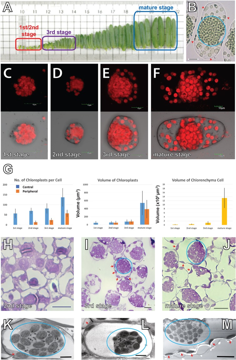
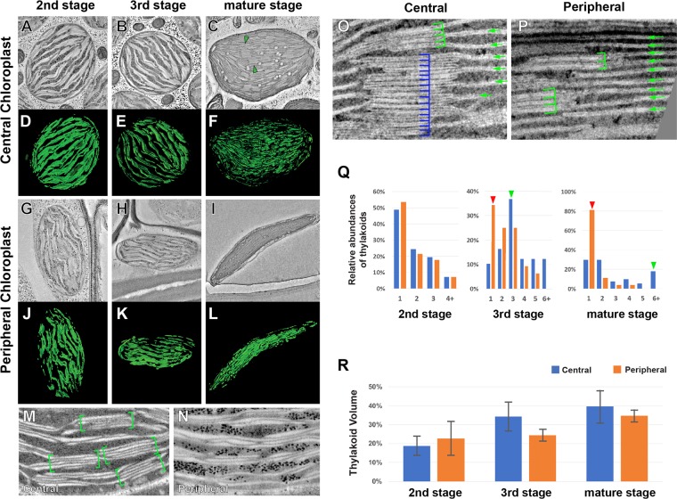
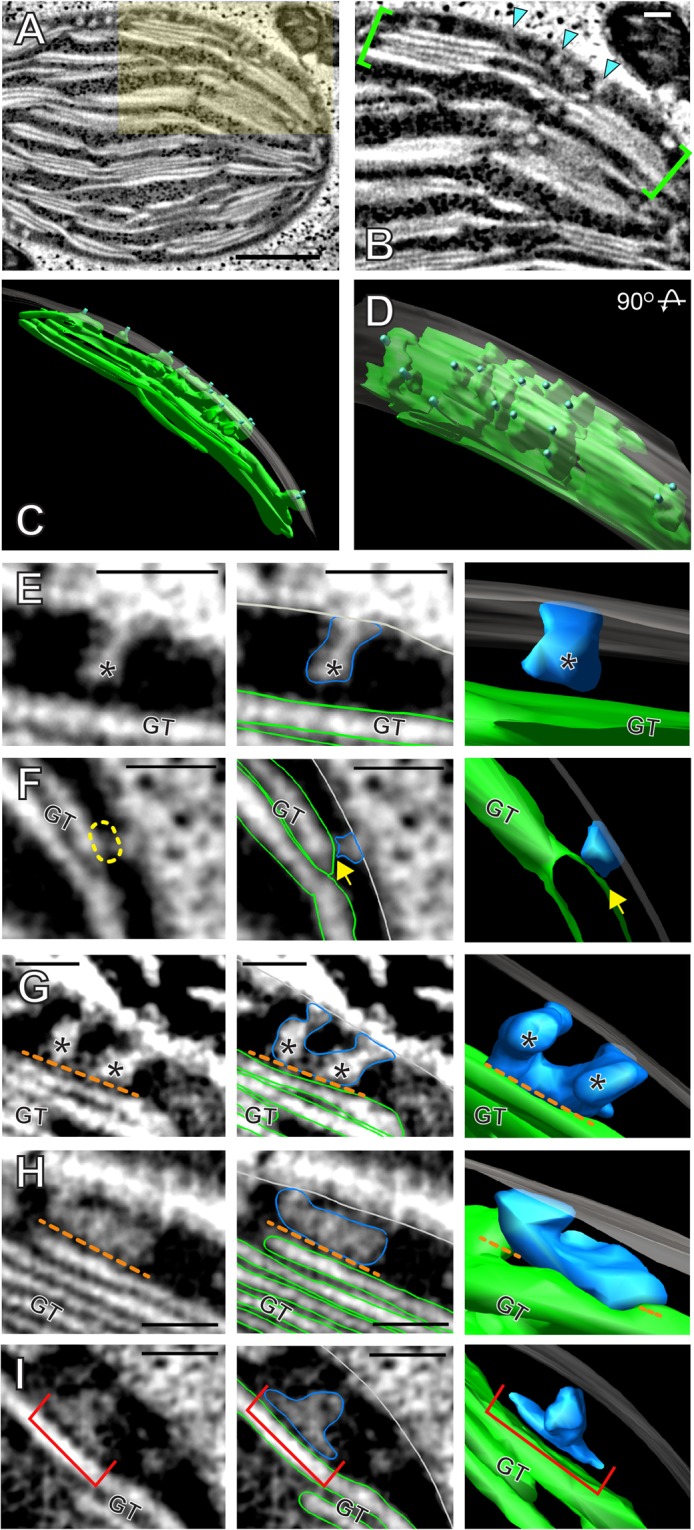
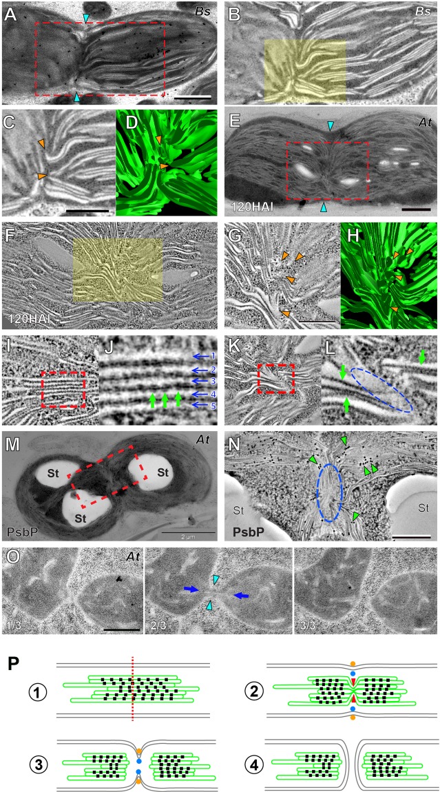
Bienertia sinuspersici is a single-cell C4 plant species of which chlorenchyma cells have two distinct groups of chloroplasts spatially segregated in the cytoplasm. The central vacuole encloses most chloroplasts at the cell center and confines the rest of the chloroplasts near the plasma membrane. Young chlorenchyma cells, however, do not have large vacuoles and their chloroplasts are homogenous. Therefore, maturing Bienertia chlorenchyma cells provide a unique opportunity to investigate chloroplast proliferation in the central cluster and the remodeling of chloroplasts that have been displaced by the vacuole to the cell periphery. Chloroplast numbers and sizes increased, more notably, during later stages of maturation than the early stages. Electron tomography analyses indicated that chloroplast enlargement is sustained by thylakoid growth and that invaginations from the inner envelope membrane contributed to thylakoid assembly. Grana stacks acquired more layers, differentiating them from stroma thylakoids as central chloroplasts matured. In peripheral chloroplasts, however, grana stacks stretched out to a degree that the distinction between grana stacks and stroma thylakoids was obscured. In central chloroplasts undergoing division, thylakoids inside the cleavage furrow were kinked and severed. Grana stacks in the division zone were disrupted, and large complexes in their membranes were dislocated, suggesting the existence of a thylakoid fission machinery.


Conflict of interest statement

The authors declare no competing interests.

Figures

Figure 1
Figure 1
Chloroplast organization in the Bienertia sinuspersici chlorenchyma cells. (A) Arrangement of Bienertia leaves according to their ages from young (left) to old (right). Sources of stages 1, 2, 3, and mature cells are indicated. The 1st stage and the 2nd stage cells are distinguishable only under the microscopes. (B) A brightfield light micrograph of a mature chlorenchyma cell. Its CC cluster (blue circle) and PCs (red arrowheads) are marked. Scale bar = 20 µm. (C–F) Confocal micrographs of chlorenchyma cells showing chlorophyll autofluorescence at (C) 1st stage, (D) 2nd stage, (E) 3rd stage, and (F) mature stage. Scale bars = 10 μm. (G) Chloroplast counts (left) and chloroplast sizes (middle) measured from chloroplast autofluorescence. Chlorenchyma cell volumes (right) were estimated from brightfield light micrographs. (H–J) Light micrographs of chlorenchyma cells from (H) 2nd stage, (I) 3rd stage, and (J) mature stage cells stained with toluidine blue. Clusters of CCs are denoted with blue circles and PCs are indicated with red arrows in. (I,J) Scale bars = 10 μm. (K–M) Transmission electron micrographs of chlorenchyma cells at (K) 2nd stage, (L) 3rd stage, and (M) mature stage. Small black dots/ovoids in the central cytoplasm inside the blue ovals correspond to mitochondria. Scale bars = 2 µm in panels K and L and 10 µm in panel M.
Figure 2
Figure 2
Electron tomography analyses of chloroplast in Bienertia chlorenchyma cells. (A–F) ET slices of central chloroplasts at (A) 2nd stage, (B) 3rd stage, (C) and mature stage. 3D models of (D) 2nd stage, (E) 3rd stage, (F) and mature stage thylakoids. Grana stacks are marked with green triangles in panel C. (G–L) ET slices of peripheral chloroplasts at (G) 2nd stage, (H) 3rd stage, and (I) mature stage. (J–L) Thylakoids from (J) 2nd stage, (K) 3rd stage, (L) and mature stage chloroplasts were rendered into 3D models. (M,N) ET slice images of a central (M) and a peripheral (N) chloroplast in 3rd stage cells. (O) Image of CC chloroplasts in mature stage cell. The number of layers in a stack varies significantly (4 for the upper stack, green brackets, and 15 for the lower stack, blue brackets). Stacks are interconnected by unstacked stroma thylakoids (green arrows). (P) Image of PC chloroplasts. Stacked and unstacked thylakoids are marked with green brackets and arrows, respectively. (Q) Abundances of thylakoids determined by number of layers per stack counted in ET slices of Bienertia chloroplasts. The difference in grana stack thickness becomes more pronounced in 3rd stage and mature stage chloroplasts. (R) Relative volumes occupied by thylakoids in Bienertia chloroplasts. The thylakoid volumes were calculated from tomographic models (n = 3) and normalized to stroma volumes of the chloroplasts.
Figure 3
Figure 3
Invaginations from the inner envelope membrane contribute to the growth of thylakoids. (A) Electron tomographic slice from a 3rd stage CC. Scale bar = 500 nm. (B) The highlighted area in panel A at a higher magnification. Buds and invaginations protrude from the inner envelope membrane (IEM) into the stromal space (cyan arrow heads). Scale bar = 100 nm. (C,D) 3D model of the bracketed region in panel B. Panel D shows the model in panel C after 90° rotation. The envelope membrane was rendered semi-transparent. (E–H) In these panels, left to right are a tomographic slice; the same slice with IEM outlined in light grey, membrane ingrowths in blue, and existing grana thylakoid (GT) in green; and a 3D model from the tomogram showing (E) an ingrowth bud emerging from the IEM. (F) An ingrowth appears to be in contact with a GT surface (yellow dashed circle / yellow arrow). (G) Two buds of similar sizes that seem to have fused (the orange dotted line marks a gap between the membranes). (H) An “L” shaped invagination that lies above an existing grana thylakoid as a separate layer (the orange dotted line marks a gap between the membranes). (H) A “T” shaped compartment (red bracket) appears to be an invagination that have separated from IEM. Scale bars = 100 nm.
Figure 4
Figure 4
Dividing chloroplasts in Bienertia and Arabidopsis cells. (A) TEM micrograph of a dividing chloroplast in a 3rd stage Bienertia cell. The cyan arrows indicate the division furrow. Scale bar = 500 nm. (B) ET slice of the boxed area in panel A. (C) Magnification of the highlighted area in panel B. The thylakoids have lost the stacked architecture and have been severed (orange arrow heads). (D) 3D model of the thylakoids shown in panel C. (E) TEM micrograph of a dividing chloroplast in an Arabidopsis cotyledon cell at 120 HAI. The cyan arrows indicate the division furrow. Scale bar = 1 µm. (F) ET slice of the boxed area in panel E. (G) Magnification of the highlighted area in panel F. The thylakoid membranes are sharply kinked (orange arrow heads) in contrast to the planar/stacked thylakoids outside the constricted zone. (H) 3D model of the thylakoids in panel G. (I,J) ET slice images of thylakoid membranes. Five lamellae of the granum in the boxed area in panel I are indicated by arrows numbered 1–5 in panel J. Pairs of electron dense complexes can be detected on opposite sides of the thylakoid membranes (green arrows). (K,L) ET slice images of twisted thylakoids in a dividing chloroplast. Panel L is the high magnification image of the boxed area in panel K. Note the absence of electron-dense complexes in the kinked region (blue dashed oval) as compared to stacked regions around it (green arrows). (M,N) Electron micrograph of a dividing Arabidopsis chloroplast. PsbP, a subunit of PSII, was localized by immunogold labeling. Panel N is a higher magnification of the boxed area of panel M showing immunogold particles (green arrow heads, 15 nm gold particles). The particles are absent in the squeezed region (blue oval). St: starch particle. Scale bars in N = 500 nm (O) Three consecutive TEM sections (1/3–3/3) of an Arabidopsis meristem cell showing a dividing proplastid. The two daughter plastids are joined via a narrow isthmus (cyan arrowheads in the middle panel). A thin tubule is seen through it (blue arrows). Scale bar = 500 nm. (P) Model of the thylakoid fission [1] A chloroplast ready to divide; indicated are inner and outer envelope membranes (grey), thylakoid membranes (green), photosystem II (PSII) complexes (black squares), and the plane of division (red dotted line). [2] The thylakoid membranes are rearranged as they are ‘pinched’ along the plane of division (red arrowheads) as seen in E and F. PSII complexes are dislocated from the plane of division. The inner (blue dots) and outer (orange dots) division rings begin squeezing the envelope membranes. [3] The thylakoid severing is complete before the division rings separate the chloroplast as seen in (A,B). [5] Two daughter chloroplasts are formed from the division of the mother chloroplast.

Similar articles

Cited by

References

    1. Gowik U, Westhoff P. The path from C3 to C4 photosynthesis. Plant Physiology. 2011;155:56–63. doi: 10.1104/pp.110.165308. - DOI - PMC - PubMed
    1. Edwards GE, Franceschi VR, Voznesenskaya EV. Single-cell C(4) photosynthesis versus the dual-cell (Kranz) paradigm. Annual Review of Plant Biology. 2004;55:173–196. doi: 10.1146/annurev.arplant.55.031903.141725. - DOI - PubMed
    1. Voznesenskaya EV, Franceschi VR, Pyankov VI, Edwards GE. Anatomy, chloroplast structure and compartmentation of enzymes relative to photosynthetic mechanisms in leaves and cotyledons of species in the tribe Salsoleae (Chenopodiaceae) J Exp Bot. 1999;50:1779–1795. doi: 10.1093/jxb/50.341.1779. - DOI
    1. Voznesenskaya EV, et al. Proof of C4 photosynthesis without Kranz anatomy in Bienertia cycloptera (Chenopodiaceae) Plant J. 2002;31:649–662. doi: 10.1046/j.1365-313X.2002.01385.x. - DOI - PubMed
    1. Sharpe RM, Offermann S. One decade after the discovery of single-cell C4 species in terrestrial plants: what did we learn about the minimal requirements of C4 photosynthesis? Photosynthesis Research. 2013;119:169–180. doi: 10.1007/s11120-013-9810-9. - DOI - PubMed

Publication types

MeSH terms