. 2020 Jan;26(1):118-130.
doi: 10.1038/s41591-019-0674-1. Epub 2019 Dec 23.

Spinal subpial delivery of AAV9 enables widespread gene silencing and blocks motoneuron degeneration in ALS

Affiliations

Spinal subpial delivery of AAV9 enables widespread gene silencing and blocks motoneuron degeneration in ALS

Mariana Bravo-Hernandez et al. Nat Med. 2020 Jan.

Abstract

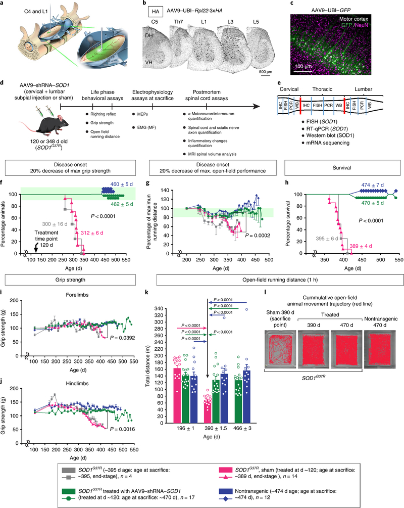
Gene silencing with virally delivered shRNA represents a promising approach for treatment of inherited neurodegenerative disorders. In the present study we develop a subpial technique, which we show in adult animals successfully delivers adeno-associated virus (AAV) throughout the cervical, thoracic and lumbar spinal cord, as well as brain motor centers. One-time injection at cervical and lumbar levels just before disease onset in mice expressing a familial amyotrophic lateral sclerosis (ALS)-causing mutant SOD1 produces long-term suppression of motoneuron disease, including near-complete preservation of spinal α-motoneurons and muscle innervation. Treatment after disease onset potently blocks progression of disease and further α-motoneuron degeneration. A single subpial AAV9 injection in adult pigs or non-human primates using a newly designed device produces homogeneous delivery throughout the cervical spinal cord white and gray matter and brain motor centers. Thus, spinal subpial delivery in adult animals is highly effective for AAV-mediated gene delivery throughout the spinal cord and supraspinal motor centers.


Conflict of interest statement

Competing interests

M. Marsala is the scientific founder of Neurgain Technologies, Inc. and has an equity interest in the company. In addition, he serves as a consultant to Neurgain Technologies, Inc., and receives compensation for these services. The terms of this arrangement have been reviewed and approved by the University of California San Diego in accordance with its policies on conflict of interests. B.K.K. is a Chief Scientific Officer in Avexis Inc. All other authors declare that no competing interests exist.

Figures

Extended Data Fig. 1 |
Extended Data Fig. 1 |. Potent Rpl22 and GFP protein expression throughout the spinal cord and brain motor centers after subpial AAV9-mediated delivery in adult wild-type (C57BL/6) mice.
a-c, Wide-spread Rpl22 protein expression in cervical, thoracic and lumbar spinal cord at 24 h after a combined cervical-C4 (10 μl) and lumbar–L1 (10 μl) AAV9-UBI-Rpl22-3xHA delivery. Sections were stained with the anti-HA antibody (black signal). d-h, An intense retrograde transduction-induced GFP expression in (d,e) nucleus ruber and (f-h) motor cortex at 14 days after a combined cervical (10 μl) and lumbar (10 μl) AAV9-UBI-GFP injection. A representative images from at least 4 individual animals are shown. ML, molecular layer.
Extended Data Fig. 2 |
Extended Data Fig. 2 |. Potent gene expression or suppression of mutant SOD1G37R expression in spinal parenchyma after spinal subpial, but not intrathecal, AAV9 delivery.
a-e, GFP expression after (a,b) intrathecal or (c-e) subpial injection of AAV-UBI-GFP. a,b, Only some α-motoneurons and occasional neurons in brain stem show GFP positivity following intrathecal injection. c-e, GFP expression throughout the entire length of spinal cord, spinocerebellar tract terminals (SCT) and brain motor centers (reticular formation-RF, nucleus ruber-NR, and motor cortex-MC) in animals subpially-injected with AAV9-UBI-GFP at single cervical (C4) and lumbar (L1) sites. e, Longitudinal spinal cord section demonstrating expression of GFP throughout the spinal cord after subpial AAV9-UBI-GFP delivery. A representative images from at least 3 individual animals per experimental group are shown. f, g, Mutant SOD1G37R RNA levels measured by FISH or Q-PCR (four weeks after AAV injections) in the lumbar spinal cord in SOD1G37R mice injected subpially or intrathecally with AAV9-shRNA-SOD1. Highly potent (over 80%) suppression of SOD1G37R mRNA in subpially-vector-injected animals can be seen with FISH and was measured with Q-PCR. No significant silencing effect in intrathecally-injected animals was detected. (f) A representative images from at least 3 individual animals per group are shown. Data are expressed as mean ± S.E.M. (SOD1G37R, n = 4; Subpial SOD1G37R AAV9-shRNA-SOD1-treated, Intrathecal SOD1G37R AAV9-shRNA-SOD1-treated and wild-type nontransgenic, n = 3 per group). Each dot represents an individual animal. Statistical significance was determined with one way ANOVA followed by Bonferroni post hoc test (ANOVA < 0.0001, F = 338.6). P values are shown between the indicated groups.
Extended Data Fig. 3 |
Extended Data Fig. 3 |. Preservation of neuromuscular junctions (NMJs) in gastrocnemius muscle in SOD1G37R mice treated before or after disease onset with AAV9-shRNA-SOD1.
a-c, Several normally-appearing NMJs triple-stained with BTX/SYN/NF-H and which are similar to (a) wilde-type nontransgenic animals can be seen in (c) SOD1G37R animals treated with AAV9-shRNA-SOD1 at age of 120 days and analyzed at age ~470 days. b, Denervated (BTX + /SYN +, but NF-H negative) NMJs are found in sham-operated SOD1G37R animals. d,e, Compared to wild-type nontransgenic animal (d) a partial denervation (loss of BTX + /SYN + /NF-H + ) is seen in SOD1G37R animals at age of ~348 days (e). f,g, Compared to end stage sham-operated animals which show extensive loss of NMJs (f) the innervation is maintained in SOD1G37R animals treated after disease onset (at age ~348 days) with AAV9-shRNA-SOD1 and analyzed at age between 398–465 days (g). A representative images from at least 3 individual animals per experimental group are shown.
Extended Data Fig. 4 |
Extended Data Fig. 4 |. Neuronal protection in cervical, thoracic and lumbar spinal cord of SOD1G37R mice after pre-symptomatic subpial injection of AAV9-shRNA-SOD1.
a,b,c, Transverse spinal cord sections taken from the cervical, thoracic and lumbar spinal cord from four sham-operated SOD1G37R animals, five SOD1G37R animals subpially-injected presymptomatically at ~120 days of age with AAV-9-shRNA-SOD1 and one non-transgenic mouse. Each mouse was analyzed between 394–474 days of age. Neurons were visualized with NeuN antibody. A clear loss of large α-motoneurons in the ventral horn (red dotted area; a, lumbar images) was seen in all four sham-operated animals at all segmental levels. NeuN staining intensity was decreased in the intermediate zone (Lamina VII; green dotted area; a, thoracic image) throughout the whole spinal cord, while there was complete preservation of α-motoneurons and NeuN staining intensity (similar to wil-type nontransgenic; (c)) in Lamina VII of (b) all five AAV9-shRNA-SOD1-treated SOD1G37R animals in cervical, thoracic and lumbar spinal cord sections.
Extended Data Fig. 5 |
Extended Data Fig. 5 |. Preservation of normal spinal mRNA profile in SOD1G37R mice treated subpially before disease onset with AAV9-shRNA-SOD1.
a, Heat map showing all genes with greater than two-fold upregulation or downregulation in all experimental groups. Expression is shown on a normalized scale from 0–1. b, Scatter plot showing differential gene expression between SOD1G37R, sham-operated SOD1G37R, wild-type nontransgenic, and SOD1G37R AAV9-shRNA-SOD1-treated mice. (gray) genes not differentially expressed; (black) genes that were differentially expressed in SOD1G37R control (end stage) + sham-operated SOD1G37R (end stage) and which expression levels were corrected in SOD1G37R mice after AAV9-shRNA-SOD1 treatment; (blue) genes still differentially expressed in SOD1G37R mice treated with AAV9-shRNA-SOD1; (magenta) partially corrected genes that are still differentially expressed in AAV9-shRNA-SOD1-treated SOD1G37R mice, but partially corrected relative to wild-type nontransgenic mice and either SOD1G37R (end stage) or sham-operated SOD1G37R (end stage). c, Clustering dendrogram demonstrating variation in gene expression across all samples. AAV9-shRNA-SOD1-treated SOD1G37R samples cluster together with wild-type nontransgenic controls and are very distinct from untreated and sham-operated SOD1G37R samples. The primary branch at the top of the dendrogram represents disease/treatment status. d, Heat map showing significant upregulation of microglial and astrocytic genes and down regulation of neuronal genes in SOD1G37R (end stage) and sham-operated SOD1G37R (end stage) mice versus wild-type nontransgenic and AAV9-shRNA-SOD1-treated SOD1G37R. e, Quantification of genes that are differentially expressed in untreated SOD1G37R (end stage) and sham-operated SOD1G37R (end stage) versus wild-type nontransgenic mice, and the number of genes that are corrected or partially corrected in AAV9-shRNA-SOD1-treated SOD1G37R mice. Number of animals used (n = number of biologically independent animals) in mRNA sequencing analysis shown above: wild-type nontransgenic (n = 4), untreated SOD1G37R end-stage disease (n = 3), sham-treated SOD1G37R end-stage disease (n = 4), presymptomatically AAV9-shRNA-SOD1-treated SOD1G37R (n = 4). Differential expression was performed using edgeR two-sided binomial test with Benjamini & Hochberg post-hoc correction.
Extended Data Fig. 6 |
Extended Data Fig. 6 |. Preservation of the remaining lumbar α-motoneurons, interneurons, and suppression of spinal parenchymal misfolded SOD1 in SOD1G37R mice treated after disease onset by subpial injection of AAV9-shRNA-SOD1.
a-d, Representative transverse lumbar spinal cord sections stained with NeuN (green) and B8H10 (magenta) antibody in mice that were (a) 489 ± 6 days old wild-type nontransgenic, (b) 348 ± 2 days old SOD1G37R, (c,d) 404 ± 14 days old, end stage SOD1G37R injected subpially with (c) AAV9-scrambled virus or (d) AAV9-shRNA-SOD1 and then assayed 90 days later at ages between 398 and 465 days. (b) α-motoneurons and early accumulation of aggregates of mutant SOD1 are seen in 348 ± 2 day old SOD1G37R mice, while those neurons and NeuN staining intensity are (c) nearly completely lost by end-stage accompanied by marked accumulation of mutant SOD1 in intermediate zone and in ventral horn. d, Remaining α-motoneurons in AAV9-shRNA-SOD1-treated SOD1G37R mice and potent suppression of aggregates of misfolded SOD1 in the ventral horn. e-h, Quantitative analysis of α-motoneuron survival, NeuN staining intensity, and misfolded SOD1 protein accumulation. Each dot represents an individual animal; at least 4 sections per animal were used. i-t, Representative transverse lumbar spinal cord sections stained with NeuN (green), GFAP (magenta), Iba1 (green) or vimentin (blue) antibodies in (i) 348 ± 2 day old wild-type nontransgenic, (j) 348 ± 2 day old SOD1G37R, and (k,l) SOD1G37R subpially injected at ~348 days of age with (k) AAV9-scrambled virus and harvested at 404 ± 14 days of age (end-stage) or (l) AAV9-shRNA-SOD1 and subsequently aged to 398–465 days (~90 days post-treatment). u-w, Quantitation of GFAP, Iba1, and vimentin in each genotype/experimental group. Each dot represents an individual animal; at least 4 sections per animal were used. Experimental animals used: SOD1G37R (348 ± 2 days of age, n = 3); Sham-operated (or injected with scramble-AAV9) SOD1G37R (n = 4); AAV9-shRNA-SOD1-treated SOD1G37R (n = 4); and wild-type nontransgenic (n = 4) mice. Data are mean ± S.E.M. Statistical significance was determined with (e) two tail unpaired t-test with Welch’s correction (P = 0.0150, t = 4.680, df = 3.288) and with (f-h, u-w) one-way ANOVA followed by Bonferroni post hoc test (f: ANOVA, P = 0.0061, F = 10.29; g: ANOVA, P = 0.0009, F = 11.71; h: ANOVA, P = 0.0394, F = 4.980; u: ANOVA, P = 0.0060, F = 7.223; v: ANOVA, P < 0.0001, F = 20.73; w: ANOVA, P = 0.0004, F = 14.30). P values are shown between the indicated groups. DH, dorsal horn; VH, ventral horn.
Extended Data Fig. 7 |
Extended Data Fig. 7 |. Preservation of lumbar spinal cord mRNA profile in SOD1G37R mice treated subpially after disease onset with AAV9-shRNA-SOD1.
a, Scatter plot showing differentially expressed genes identified (by RNA seq) between non-treated SOD1G37R animals analyzed after disease onset (~348 days of age) and untreated end-stage SOD1G37R animals (~395 days of age). Compared to comparable analyses from mid-stage disease animals, 593 genes were upregulated and 685 downregulated in end-stage SOD1G37R mice. b, Scatter plot showing differentially expressed genes between non-treated SOD1G37R animals after disease onset (~348 days of age) and SOD1G37R animals treated with AAV9-shRNA-SOD1 after disease onset (at ~348 days of age) and surviving for additional 96 days with no clinical signs of disease. Only 47 genes were upregulated and 66 downregulated when comparing mid-stage diseased animals to AAV9-shRNA-SOD1-treated SOD1G37R mice. Each dot in a and b represents one gene. Black circles represent genes that are differentially expressed between the two conditions, and gray circles represent genes that are statistically unchanged. Open triangles represent genes that fall outside of the y-axis scale (only two genes fit this category in A). c, Minimum spanning tree plots sampled by expression of SOD1 disease-associated genes generated using DDRTree dimension reduction in Monocle. Data from all experimental groups are presented. Wild-type nontransgenic mice and SOD1G37R mice treated with AAV9-shRNA-SOD1 before disease onset (at age~120 days) cluster to the left; mid-stage disease SOD1G37R, non-treated and SOD1G37R treated at mid-stage disease (at age ~348 days) and then surviving for additional 96 days cluster in the middle. All non-treated end-stage (end stage disease) SOD1G37R animals cluster at the right. Number of animals used (n = number of biologically independent animals) in mRNA sequencing analysis shown above: wild-type nontransgenic (n = 5), untreated SOD1G37R end-stage disease (n = 3), sham-treated SOD1G37R end-stage disease (n = 4), presymptomatic AAV9-shRNA-SOD1-treated SOD1G37R (n = 4), untreated SOD1G37R mid-stage disease (n = 3), postsymptomatic AAV9-shRNA-SOD1-treated SOD1G37R (n = 3). Differential expression was performed using edgeR two-sided binomial test with Benjamini & Hochberg post-hoc correction.
Extended Data Fig. 8 |
Extended Data Fig. 8 |. Widespread GFP expression in pyramidal neurons of motor cortex after a single bolus cervical subpial delivery of AAV9-UBI-GFP in adult pig.
a-f, Intense GFP expression throughout the majority of pyramidal neuron population in layer V of the motor cortex at 3 weeks after a single bolus cervical subpial delivery of AAV9-UBI-GFP in an adult pig. GFP expression was not detected in glial cells localized in the molecular layer, consistent with retrograde AAV delivery to pyramidal neurons from corticospinal axons transiting through or terminating in subpially-injected cervical spinal cord segments. A representative result of 3 individual pigs is shown. ML, molecular layer.
Extended Data Fig. 9 |
Extended Data Fig. 9 |. Limited GFP expression in motor cortex neurons after single intrathecal bolus cervical delivery of AAV9-UBI-GFP to adult pig.
a-e, Irregularly distributed GFP+ immunoreactive regions in the cortical molecular layer, with GFP+ neurons in layer II-III (d, white-boxed area) 21 days following single intrathecal bolus cervical delivery of AAV9-UBI-GFP in an adult pig. f, g, GFP+ non-pyramidal neurons identified in deeper cortical layers, primarily found in the vicinity of cerebral arteries, suggesting perivascular AAV9 diffusion into brain parenchyma. A representative result of 3 individual pigs is shown. Art, arterial lumen.
Extended Data Fig. 10 |
Extended Data Fig. 10 |. Effective AAV9-encoded gene expression throughout the entire cervical spinal cord of the adult monkey after a single bolus, subpial C3 delivery of AAV9-UBI-Rpl22-3×HA.
a-c, An intraoperative photograph depicting the placement of subpial injection needle in the C3-C4 segment and rostro-caudal spread of ‘blue’ dextran (10,000 MW; 300 μl) immediately after initiation of subpial infusion (1 μl/5 sec). d-f, Expression of Rpl22 protein (anti-HA staining; green signal) in cervical spinal cord segments (C1-C8) at 48 h after mid-cervical subpial AAV9-UBI-Rpl22-3xHA delivery. g-l, Neuronal (NeuN), astrocyte (GFAP) and oligodendrocyte (Olig2)-expression of Rpl22 protein (HA+ signal) at 48 h after AAV9-UBI-Rpl22-3xHA delivery. m, n, Absence of specific staining in non-injected animals. A representative result of 2 individual non-human primates per group is shown (Subpially-injected, n = 2; Control non-injected, n = 2). DH, dorsal horn; VH, ventral horn.
Fig. 1 |
Fig. 1 |. Spinal SP AAV9–shRNA–SOD1 delivery before disease onset in SOD1G37R mice blocks disease onset, producing long-term preservation of motor function.
a, Schematic of upper cervical and lumbar SP delivery of AAV9. b,c Widespread AAV9 delivery and expression of an AAV9-encoded gene in the dorsal and ventral horns of adult mice, identified by immunohistochemistry of spinal cord sections taken from different segmental levels (b) or immunofluorescence of motor cortex (c) after a single upper cervical and lumbar SP delivery of AAV9–Rpl22-x3HA (b) (imaged 48 h after AAV9 injection) or AAV9–UBI–GFP (c) (imaged 14 d after AAV9 injection), retrogradely delivered to the motor cortex. A representative result from four independent mice is shown. d,e, Schematic diagrams of the experimental design and ‘in-life’ and ‘postmortem’ assays. f,g, Disease onset (defined as 20% decrease of maximum grip strength (f) or 20% decrease of maximum open-field performance (g)). h, Survival (measured by normal righting reflex) in SOD1G37R mice with or without SP AAV9–shRNA–SOD1 injection before disease onset. i,j, Grip strength in forelimbs (i) and hindlimbs (j) with and without AAV9–shRNA–SOD1 treatment. k,l, Open-field motor performance (running distance per 1 h) with and without SP AAV9–shRNA–SOD1 treatment. Data are expressed as mean ± s.e.m. (squares: nontreated SOD1G37R mice (n = 4); triangle: sham-operated SOD1G37R mice (n = 14); circles: shRNA–SOD1-treated SOD1G37R mice (n = 17); diamond: normal wild-type mice (n = 12)). The statistical significance was determined as follows: Kaplan–Meier survival analysis using the two-sided log-rank test (f,h); two-way ANOVA followed by Bonferroni’s posthoc test (g: ANOVA Pint = 0.8737, F = 0.7800; i, ANOVA, Pint < 0.0001, F = 1.763; j: ANOVA Pint < 0.0001, F = 3.50); k, Kruskal–Wallis test followed by Steel–Dwass posthoc test (P < 0.0001). P values are shown between the indicated groups/time points. DH, dorsal horn; VH, ventral horn.
Fig. 2 |
Fig. 2 |. Continuing presence of myogenic MEPs and lack of MF in SOD1G37R mice treated before disease onset with AAV9–shRNA–SOD1.
a,b, Schematics of MEPs (a) and EMG (muscle fibrillation: MF) (b) recordings. c,d, Representative MEP recordings from three individual wild-type nontransgenic (left), shamo-perated SOD1G37R (middle) and AAV9–shRNA–SOD1-treated SOD1G37R mice (right). A complete loss of MEP response is seen in sham-operated SOD1G37R mice (c, middle panel boxes), whereas AAV9–shRNA–SOD1-treated SOD1G37R mice showed continued MEPs (c, right panel boxes), with notable preservation of amplitude (d). e,f, Representative MF recordings from three wild-type nontransgenic mice (left), sham-operated SOD1G37R mice (middle) and AAV9–shRNA-treated SOD1G37R mice (right) mice. g–j, Microphotographs and axonal quantification of lateral funiculi (Th10) (g,h) or sciatic nerves (i,j) of wild-type nontransgenic, untreated SOD1G37R or AAV9–shRNA–SOD1-treated SOD1G37R mice. Data for MEPs (d) and MF (f) are expressed as mean ± s.e.m.; each dot represents an individual animal. One-way ANOVA was used for statistical analyses (d: wild-type nontransgenic mice, n = 3; sham-operated SOD1G37R mice, n = 4; shRNA–SOD1-treated SODG37R mice, n = 8; P = 0.0007, F = 14.09 using the Holm–Sidak posthoc test; f: wild-type nontransgenic mice, n = 4; sham-operated SOD1G37R mice, n = 17; shRNA–SOD1-treated SODG37R mice, n = 14; P < 0.0001, F = 90.55 using the Holm–Sidak posthoc test). Data on axonal counts (h,j) are expressed as mean ± s.e.m. (wild-type nontransgenic mice, n = 3; SOD1G37R nontreated mice, n = 4; shRNA–SOD1-treated SODG37R mice, n = 3). The statistical significance was determined using one-way ANOVA followed by Bonferroni’s posthoc test (h, 2–4 μm: ANOVA, P = 0.0020, F = 17.19, h, >4 μm: ANOVA P = 0.0030, F = 14.83; j, 0–4 μm: ANOVA,P = 0.0076, F = 10.60, j, >4 μm: ANOVA P = 0.0007, F = 23.98). The P values are shown between the indicated groups.
Fig. 3 |
Fig. 3 |. Preservation of spinal α-motoneurons and interneurons, and suppression of misfolded SOD1 protein accumulation in spinal parenchyma of AAV9–shRNA–SOD1-treated SOD1G37R mice treated before disease onset.
a–c, NeuN, misfolded SOD1 (identified by the B8H10 antibody25) or choline acetyltransferase in lumbar spinal cord sections of wild-type nontransgenic mice (a) or SOD1G37R mice without (b) or with (c) SP treatment with AAV9–shRNA–SOD1 before disease onset. A representative result of at least 12 individual sham-operated SOD1G37R mice, 8 individual AAV9–shRNA–SOD1-treated SOD1G37R mice and 3 individual wild-type nontransgenic mice is shown. df, Loss of lumbar interneurons expressing NeuN between laminae IV and VII in sham-operated SOD1G37R mice (e) relative to wild-type nontransgenic (d) but not after treatment with AAV9–shRNA–SOD1 (f) before disease onset. A representative result of 13 individual sham-operated SOD1G37R mice, 8 AAV9–shRNA–SOD1-treated SOD1G37Rmice and 4 wild-type nontransgenic mice is shown. gi, Quantitative analysis of cervical, thoracic and lumbar α-motoneurons, NeuN expression (lumbar spinal cord) and accumulation of misfolded SOD1 in the cervical and lumbar spinal cord of wild-type nontransgenic, AAV9–shRNA–SOD1-treated SOD1G37R and untreated SOD1G37R mice using at least four sections per animal. All data are expressed as mean ± s.e.m. Each circle represents an individual mouse. Sham-operated SOD1G37R mice (n ≥ 12), AAV9–shRNA–SOD1-treated SOD1G37R mice (n ≥ 8) and wild-type nontransgenic mice (n ≥ 3); the exact individual sample sizes are shown in the corresponding graph. One-way ANOVA followed by Bonferroni’s posthoc test was used for statistical analysis in g and h (g, cervical: ANOVA P < 0.0001, F = 100.6, g, thoracic: ANOVA P < 0.0001, F = 47.69; g, lumbar: ANOVA P < 0.0001, F = 134.5; h, ANOVA P < 0.0001, F = 62.16). The unpaired, two-tailed, Student’s t-test with Welch’s correction was used in i (i, cervical: P = 0.0202, t = 2.534, degrees of freedom (d.f.) = 19.04; i, lumbar: P = 0.0001, t = 5.354, d.f. = 13.63.) P values are shown between the indicated groups, DH, dorsal horn; VH, ventral horn.
Fig. 4 |
Fig. 4 |. Blockage of spinal cord atrophy and suppression of inflammatory changes and mutant SOD1 mRNA and protein accumulation in SOD1G37R mice treated before disease onset with AAV9–shRNA–SOD1.
a,b, Notable preservation of the gray matter volume and the gray:white matter ratio determined by MRI-based volumetric analysis of lumbar enlargement in SOD1G37R mice treated before disease onset with AAV9–shRNA–SOD1. Representative images (a) and the result of quantification and statistical analysis (b) are shown. Each dot represents an individual animal. ch, Suppression of astrocyte activation (marked by GFAP) (ce) and microglial activation (marked by Iba1) (fh) in SOD1G37R mice treated before disease onset with AAV9–shRNA–SOD1, relative to areas of α-motoneuronal loss and decreased NeuN+ staining in the intermediate zone, identified in transverse lumbar sections in sham-operated SOD1G37R mice. i,j, Quantification by densitometry of Iba 1 (i) and GFAP (j) reactivity in gray matter of wild-type nontransgenic and treated and untreated SOD1G37R mice, using at least four sections per animal. A representative typical image for each group is shown; each dot represents an individual animal. ks, Mutant SOD1 RNA levels (measured with FISH) in cervical, thoracic and lumbar sections from wild-type nontransgenic (k,n,q), sham-operated SOD1G37R (l,o,r) and SOD1G37R (m,p,s) animals treated before disease onset with AAV9–shRNA–SOD1. A representative image of each segment for at least three animals from each group is shown. tv, Analysis of mutant SOD1 RNA or protein levels by qPCR (t) or immunoblot (u,v) of cervical, thoracic and lumbar spinal cord and liver tissue from each experimental group. Each dot represents an individual animal. Representative cropped immunoblot bands are shown for each group (see Supplementary Fig. 5 and Source data Fig. 1). All data are expressed as mean ± s.e.m. Untreated SOD1G37R mice (b: n = 4), sham-operated SOD1G37R mice (i,j: n = 12; t,u,v: n = 3), AAV9–shRNA–SOD1-treated SOD1G37R mice (b: n = 3; i,j: n = 9; t,u,v: n = 4) and wild-type nontransgenic mice (n = 3). The statistical significance was determined using one-way ANOVA followed by Bonferroni’s posthoc test (b, left graph: ANOVA P < 0.0001, F = 46.65; b, right graph: ANOVA P = 0.0024, F = 16.16; i: ANOVA P = 0.0009, F = 10.06; j: ANOVA P < 0.0001, F = 23.60; t, cervical: ANOVA P < 0.0001, F = 57.14; t, thoracic: ANOVA P < 0.0001, F = 124.8; t, lumbar: ANOVA P < 0.0001, F = 692.1; u, cervical: ANOVA P = 0.0003, F = 33.663.60; u, thoracic: ANOVA P < 0.0001, F = 147.9; u, lumbar: ANOVA P < 0.0001, F = 48.15; v: ANOVA P < 0.0001, F = 93.37). P values are shown between the indicated groups. DH, dorsal horn; VH, ventral horn.
Fig. 5 |
Fig. 5 |. Spinal SP AAV9–shRNA–SOD1 delivery to symptomatic SOD1G37R mice blocks further disease progression and preserves residual motor function.
a, Presence of fibrillations in the GCM and corresponding α-motoneuron degeneration in 348 ± 2-d-old SOD1G37R mice. The result of three independent animals is shown. b, Baseline EMG activity (background) and normal spinal α-motoneuron morphology (visualized by immunofluorescence for NeuN) in wild-type nontransgenic age-matched (age 348 ± 2 d) mice. A representative result of four independent mice is shown. c, Quantitative analysis of MFs (integrated EMG signals) in all experimental groups. Each dot represents an individual animal. df, Disease onset, survival (measured by loss of righting reflex) and hindlimb grip strength in nontransgenic SOD1G37R and AAV9–shRNA–SOD1-treated SOD1G37R mice. g, MF recording and corresponding lumbar spinal cord α-motoneuron degeneration (blue rectangle areas) in four individual sham-operated or scrambled virus-injected SOD1G37R animals, recorded at the end-stage of disease. High-amplitude MFs and extensive α-motoneuron loss can be seen in all animals. h, MF recordings and corresponding lumbar spinal cord α-motoneuron preservation (rectangular areas) in four individual shRNA–SOD1-injected SOD1G37R animals. Note the complete absence of MF with large α-motoneuronal protection in animals nos. 254 and 234. Data are expressed as mean ± s.e.m. Untreated SOD1G37R mice (n = 3), sham or scramble and AAV9–shRNA–SOD1-treated SOD1G37R mice (n = 4 for each group), AAV9–shRNA–SOD1-treated SOD1G37R mice (n = 4) and wild-type nontransgenic mice (n = 4). All animals in g and h were sham/scramble-vector injected or treated with AAV9–shRNA–SOD1 at age 348 ± 2 d. The statistical significance was determined as follows: c, unpaired, two-tailed, Student’s t-test (d.f. = 6); d, Kaplan–Meier survival analysis using the two-sided log-rank test (there was no difference in median disease onset across SOD1G37R, sham- or scramble-vector-treated SOD1G37R mice (n = 4; 367 d) and AAV9–shRNA–SOD1-treated SOD1G37R mice (n = 4, 374 d), with an average age at treatment of 348 d, P = 0.5352; e, Kaplan–Meier survival analysis using the log-rank test: median survival of AAV9–shRNA–SOD1-treated SOD1G37R animals (n = 4, undefined; three of four animals continued to survive for a minimum of 451 d) versus SOD1G37R, sham or scramble-operated mice (n = 4, 402 d; P = 0.0510); f, two-way ANOVA followed by Bonferroni’s posthoc test (ANOVA Pint < 0.0001, F = 2.194). P values are shown between the indicated groups/time points.
Fig. 6 |
Fig. 6 |. Potent AAV9-mediated gene delivery into the cervical spinal cord and brain motor centers in adult pigs after a single-bolus SP AAV9–UBI–GFP injection.
a, Functional prototype of human SP injection device composed of a self-anchoring platform, built-in muscle retractors, spinal titanium clamps, XYZ manipulator (Narishige) and 27-G SP needle. The injector is used for SP vector delivery of AAV9–UBI–GFP (400 μl) into adult (35–45 kg) Gottingen–Minnesota pigs. b, SP injection needle just before and after placement into spinal cervical SP space. c, GFP expression in the white and gray matter of C2–C8 cervical segments (21 d after AAV9–UBI–GFP injection). dm, Retrograde transduction-induced GFP expression in tectal (e), rubral (g,i,j) reticular (h) and cortical–pyramidal (km) neurons. A representative result of three independent mini-pigs is shown.

References

    1. Rosen DR et al. Mutations in Cu/Zn superoxide dismutase gene are associated with familial amyotrophic lateral sclerosis. Nature 362, 59–62 (1993). - PubMed
    1. Matsumoto A et al. Disease progression of human SOD1 (G93A) transgenic ALS model rats. J. Neurosci. Res. 83, 119–133 (2006). - PubMed
    1. Kaur SJ, McKeown SR & Rashid S Mutant SOD1 mediated pathogenesis of amyotrophic lateral sclerosis. Gene 577, 109–118 (2016). - PubMed
    1. van Zundert B & Brown RH Jr. Silencing strategies for therapy of SOD1-mediated ALS. Neurosci. Lett. 636, 32–39 (2017). - PubMed
    1. Howland DS et al. Focal loss of the glutamate transporter EAAT2 in a transgenic rat model of SOD1 mutant-mediated amyotrophic lateral sclerosis (ALS). Proc. Natl Acad. Sci. USA 99, 1604–1609 (2002). - PMC - PubMed

Publication types

MeSH terms

Supplementary concepts