Clinical Trial
. 2020 Apr 1;130(4):1669-1682.
doi: 10.1172/JCI129301.

Distinct interferon signatures and cytokine patterns define additional systemic autoinflammatory diseases

Adriana A de Jesus  1 Yangfeng Hou  2 Stephen Brooks  3 Louise Malle  4 Angelique Biancotto  5 Yan Huang  1 Katherine R Calvo  6 Bernadette Marrero  7 Susan Moir  8 Andrew J Oler  9 Zuoming Deng  3 Gina A Montealegre Sanchez  1 Amina Ahmed  10   11 Eric Allenspach  10   12 Bita Arabshahi  10   13 Edward Behrens  10   14 Susanne Benseler  10   15 Liliana Bezrodnik  10   16 Sharon Bout-Tabaku  10   17 AnneMarie C Brescia  10   18 Diane Brown  10   19 Jon M Burnham  10   14 Maria Soledad Caldirola  10   16 Ruy Carrasco  10   20 Alice Y Chan  10   21 Rolando Cimaz  10   22 Paul Dancey  10   23 Jason Dare  10   24 Marietta DeGuzman  10   25 Victoria Dimitriades  10   26 Ian Ferguson  10   27 Polly Ferguson  10   28 Laura Finn  10   29 Marco Gattorno  10   30 Alexei A Grom  10   31 Eric P Hanson  10   32 Philip J Hashkes  10   33 Christian M Hedrich  10   34 Ronit Herzog  10   35 Gerd Horneff  10   36 Rita Jerath  10   37 Elizabeth Kessler  10   38 Hanna Kim  10   39 Daniel J Kingsbury  10   40 Ronald M Laxer  10   41 Pui Y Lee  10   42 Min Ae Lee-Kirsch  10   43 Laura Lewandowski  10   44 Suzanne Li  10   45 Vibke Lilleby  10   46 Vafa Mammadova  10   47 Lakshmi N Moorthy  10   48 Gulnara Nasrullayeva  10   47 Kathleen M O'Neil  10   32 Karen Onel  10   49 Seza Ozen  10   50 Nancy Pan  10   49 Pascal Pillet  10   51 Daniela Gp Piotto  10   52 Marilynn G Punaro  10   53 Andreas Reiff  10   54 Adam Reinhardt  10   55 Lisa G Rider  10   56 Rafael Rivas-Chacon  10   57 Tova Ronis  10   58 Angela Rösen-Wolff  10   43 Johannes Roth  10   59 Natasha Mckerran Ruth  10   60 Marite Rygg  10   61 Heinrike Schmeling  10   15 Grant Schulert  10   31 Christiaan Scott  10   62 Gisella Seminario  10   16 Andrew Shulman  10   63 Vidya Sivaraman  10   64 Mary Beth Son  10   65 Yuriy Stepanovskiy  10   66 Elizabeth Stringer  10   67 Sara Taber  10   68 Maria Teresa Terreri  10   52 Cynthia Tifft  10   69 Troy Torgerson  10   12 Laura Tosi  10   70 Annet Van Royen-Kerkhof  10   71 Theresa Wampler Muskardin  10   72 Scott W Canna  73 Raphaela Goldbach-Mansky  1
Affiliations
Clinical Trial

Distinct interferon signatures and cytokine patterns define additional systemic autoinflammatory diseases

Adriana A de Jesus et al. J Clin Invest. .

Abstract

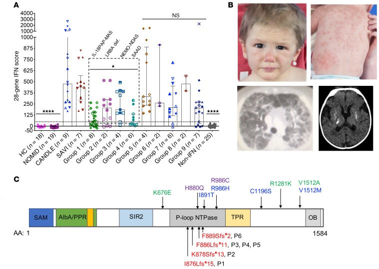
BACKGROUNDUndifferentiated systemic autoinflammatory diseases (USAIDs) present diagnostic and therapeutic challenges. Chronic interferon (IFN) signaling and cytokine dysregulation may identify diseases with available targeted treatments.METHODSSixty-six consecutively referred USAID patients underwent underwent screening for the presence of an interferon signature using a standardized type-I IFN-response-gene score (IRG-S), cytokine profiling, and genetic evaluation by next-generation sequencing.RESULTSThirty-six USAID patients (55%) had elevated IRG-S. Neutrophilic panniculitis (40% vs. 0%), basal ganglia calcifications (46% vs. 0%), interstitial lung disease (47% vs. 5%), and myositis (60% vs. 10%) were more prevalent in patients with elevated IRG-S. Moderate IRG-S elevation and highly elevated serum IL-18 distinguished 8 patients with pulmonary alveolar proteinosis (PAP) and recurrent macrophage activation syndrome (MAS). Among patients with panniculitis and progressive cytopenias, 2 patients were compound heterozygous for potentially novel LRBA mutations, 4 patients harbored potentially novel splice variants in IKBKG (which encodes NF-κB essential modulator [NEMO]), and 6 patients had de novo frameshift mutations in SAMD9L. Of additional 12 patients with elevated IRG-S and CANDLE-, SAVI- or Aicardi-Goutières syndrome-like (AGS-like) phenotypes, 5 patients carried mutations in either SAMHD1, TREX1, PSMB8, or PSMG2. Two patients had anti-MDA5 autoantibody-positive juvenile dermatomyositis, and 7 could not be classified. Patients with LRBA, IKBKG, and SAMD9L mutations showed a pattern of IRG elevation that suggests prominent NF-κB activation different from the canonical interferonopathies CANDLE, SAVI, and AGS.CONCLUSIONSIn patients with elevated IRG-S, we identified characteristic clinical features and 3 additional autoinflammatory diseases: IL-18-mediated PAP and recurrent MAS (IL-18PAP-MAS), NEMO deleted exon 5-autoinflammatory syndrome (NEMO-NDAS), and SAMD9L-associated autoinflammatory disease (SAMD9L-SAAD). The IRG-S expands the diagnostic armamentarium in evaluating USAIDs and points to different pathways regulating IRG expression.TRIAL REGISTRATIONClinicalTrials.gov NCT02974595.FUNDINGThe Intramural Research Program of the NIH, NIAID, NIAMS, and the Clinical Center.

Keywords: Genetic diseases; Immunology; Inflammation; Innate immunity; Monogenic diseases.


Conflict of interest statement

Conflict of interest: RGM received investigator-initiated grants under government collaborative agreements from SOBI, Lilly, Regeneron and Novartis. SWC received grants from Novartis and AB2Bio. JAD received grants from Pfizer, Roche, and Bristol-Myers Squibb. AAG received grants from NovImmune, grants and personal fees from Ab2Bio, and grants from Novartis. RML received consultant fees from SOBI and Novartis. LGR received research support from Hope Pharmaceuticals, Bristol-Myers Squibb, and Elli Lily. GS received personal fees from Novartis. TLWM received grants from Arthritis National Research Foundation, NYU Clinical & Translational Science Institute; personal fees from Novartis; and has a patent Methods and Materials for Treating Autoimmune Conditions issued.

Figures

Figure 1
Figure 1. Study overview, patient allocation and diagnosis.
All patients were screened for elevation of an IFN-response-gene score (IRG-S) except for 1 patient (G4-P5), who was diagnosed postmortem based on WES with SAMD9L-SAAD. Patients were characterized based on presence or absence of an elevated IRG-S. All patients underwent clinical phenotyping, cytokine analyses, and genetic testing (WES/WGS or Sanger sequencing). Negative–IRG-S patients were clinically grouped (see supplement). Positive–IRG-S patients were grouped as CANDLE-like, SAVI-like, and AGS-like disease. Cytokine analyses and genetic analyses allowed for the characterization of patients with 3 additional diseases: IL-18PAP-MAS, NEMO-NDAS, and SAMD9L-SAAD (in red); and 2 patients had LRBA deficiency. Three patients had CANDLE, 1 AGS5, and 2 had anti-MDA5 autoantibody positive juvenile dermatomyositis. Seven patients with an elevated IRG-S and 7 patients with negative IRG-S could not be further classified. *No monogenic candidate gene. **Negative–IRG-S patients were classified as CRMO/CRMO-like (n = 5), CAPS/CAPS-like (n = 4), periodic fever syndrome (n = 6), Still’s-like disease and MAS (n = 3); 7 patients could not be classified. ***Two patients (G1-P5 and G4-P6) only had one sample, 1 patient (G4-P3) had a bone marrow transplant (BMT) and no pre-BMT sample available, and 1 patient (G1-P3) had 3 negative samples.
Figure 2
Figure 2. Shared clinical features and different cytokine profiles in patients with and without elevated IFN scores.
(A) An elevated IFN-response gene score (IRG-S) distinguishes 36 patients during active disease from 29 patients who had normal IFN scores during bouts of active disease. Nonparametric (Kruskal-Wallis) test was used for multiple comparisons of IFN or Non-IFN groups with healthy controls (HC) or with CANDLE and SAVI patients combined. Depicted in the graph are the statistical significances (Kruskal-Wallis test) from the comparisons of each group (NOMID, IFN, and Non-IFN groups) with CANDLE and SAVI patients combined. Each individual patient is represented by a different symbol shape. ****P < 0.0001; NS, not significant. Bars and error lines indicate median and interquartile range, respectively; dotted line indicates the 28-gene IFN score cutoff (48.9) previously described (34). Multiple comparisons of each group (NOMID, CANDLE, and SAVI combined; IFN and Non-IFN) with HC (not depicted): NOMID P = 0.5004, CANDLE + SAVI P < 0.0001, IFN P < 0.0001, Non-IFN P = 0.2986. For HC, NOMID, and non-IFN groups, the same symbol is used for different individuals, as only 1 sample per patient is included. For CANDLE, SAVI, and IFN groups, each patient is represented by a different symbol. (BG) Characteristic clinical features that were present only in patients with elevated IFN scores included panniculitis with lipoatrophy (B), neutrophilic vasculitis (C), erythematous macular rash (D), Gottron’s papules (E), interstitial lung disease (F), and basal ganglia calcifications (G). Four patients (per groups defined in Table 2; Group 1 – patient 3 [G1-P3] and G1-P5, G4-P2 and G4-P5) had negative IRG-S but were later added to the respective groups when a clinical or genetic diagnosis was made (not depicted).
Figure 3
Figure 3. Clinical features and cytokine dysregulation in patients with IL-18PAP-MAS (n = 8).
(A) Nail clubbing. (B) Interstitial and alveolar lung disease. (C) Histologic features characteristic of pulmonary alveolar proteinosis (PAP). Original magnification, ×20. (D) Heatmap of 22 out of 48 analytes tested. The 12-cytokine signature includes SDF-1α/CXCL12, IFN-α2, MCP-3/CCL7, β-NGF, TRAIL, SCF, IL-16, IL-3, IL-18, IL-12p40, TNF-β/Ltα, and M-CSF. The 12-cytokine signature upregulation tracks with ultrahigh IL-18 levels that are also seen in patients with recurrent macrophage activation syndrome (MAS) and gain-of-function mutations in NLRC4 (12). Two other patients with the 12-cytokine signature (*) had MAS but no pulmonary disease. (E) Serum IL-18 levels in patients with IL-18PAP-MAS and healthy and disease controls. HC, healthy controls; IL-18PAP-MAS, G1 patients with PAP and recurrent MAS (n = 8); NLRC4/MAS, patients with monogenic NLRC4-mediated MAS (n = 5); NOMID, neonatal-onset multisystem inflammatory disease (n = 8); SAVI, STING-associated vasculopathy with onset in infancy (n = 5); CANDLE, chronic atypical neutrophilic dermatosis with lipodystrophy and elevated temperatures (n = 8). (F) IL-18/CXCL9 ratio has previously been described to a cutoff of 2.3 (15), which is indicated with a gray horizontal line. IL-18PAP-MAS (n = 7); NLRC4/MAS, patients with monogenic NLRC4-mediated MAS (n = 5). In E and F, nonparametric test (Kruskal-Wallis) was performed for multiple comparisons and all significant differences are shown. In E, populations in brackets share the same significance pattern. *P < 0.05; **P < 0.01; ****P < 0.0001.
Figure 4
Figure 4. IFN score by disease group and clinical and genetic characteristics of SAMD9L-mediated autoinflammatory disease (SAMD9L-SAAD).
(A) Twenty-eight–gene IFN scores by disease group. Nonparametric tests were used for multiple comparisons (Kruskal-Wallis) or individual group comparisons (Mann-Whitney). Each patient in groups 1 to 9 (G1 to G9) is represented by a different symbol shape. Depicted in the graph are the statistical significances (Kruskal-Wallis test) from the comparisons of each group (healthy controls [HC], NOMID, G1 to G4 combined, G5, G6, G7, G8, G9, and Non-IFN groups) with CANDLE and SAVI patients combined. *P = 0.0259; ****P < 0.0001; NS, not significant. Bars and error lines indicate median and interquartile range, respectively; dotted line indicates the 28-gene IFN score cutoff (48.9) previously described (34). For the control groups, HC and NOMID, the same symbol is used for different individuals, as only 1 sample per patient is included. For all other disease groups including CANDLE and SAVI, each patient is represented by a different symbol. (B) Clinical manifestations of SAMD9L-associated autoinflammatory disease (SAMD9L-SAAD) include nodular panniculitis and lipoatrophy (patient 1), interstitial lung disease (patient 6), and basal ganglia calcifications (patient 5). (C) SAMD9L domains and variant localization. SAM (blue), sterile α motif domain; AlbA/PPR (orange/yellow), DNA-binding domain; SIR2 (light blue), silent mating-type information regulator 2; P-loop NTPase (green), P-loop–containing NTP hydrolase; TPR (purple), tetratricopeptide repeat domain; OB (gray), oligonucleotide-binding fold domain; AA, amino acid; P1–P6, patient 1 to patient 6. Variants identified in SAMD9L-SAAD are in red, variants associated with ataxia-pancytopenia syndrome (APS) are in blue, and variants associated with myelodysplastic syndrome (MDS) or acute myeloid leukemia (AML) are in green. Variants associated with either APS or MDS/AML are shown in purple.
Figure 5
Figure 5. Ratios of 3-gene score (CXCL10 + GBP1 + SOCS1) over 25-gene score (3:25 gene ratio).
(A) A ratio between 3 IFN-response genes with NF-κB transcription binding sites (CXCL10, GBP1, and SOCS1) and the 25 IFN genes with no NF-κB binding sites (3:25 ratio) was calculated. Depicted in the graph are the statistical significances (nonparametric Kruskal-Wallis test) from the comparisons of each group with CANDLE and SAVI patients combined: healthy controls (HC) and NOMID combined P < 0.0001, G1 P = 0.0017, G2 P = 0.0003, G3 P < 0.0001, G4 P = 0.0027, G5 P = 0.9271, G6 P = 0.6055, G7 P = 0.1499, G8 P = 0.8684, G9 P = 0.0083, Non-IFN P < 0.0001. NS, not significant. Bars and error lines indicate median and interquartile range, respectively. Red shaded area indicates a high 3:25 gene ratio, blue shaded area indicates low ratio, and white area indicates normal ratio. Cutoffs were calculated in panel B. For the control groups (HC and NOMID) the same symbol is used for different individuals, as only one sample per patient is included. In all other groups, including CANDLE and SAVI, each patient is represented by a different symbol. (B) Two receiver operating characteristic (ROC) curves for the 3:25 gene ratio to distinguish HC and NOMID from patients with NDAS (G3) and LRBA deficiency (G2) (ROC-1) and CANDLE and SAVI from HC and NOMID (ROC-2) are shown. A black arrow indicates the optimal cutoff (listed under each graph). AUC, area under the curve; SEN, sensitivity; SPE, specificity. The cutoffs for the ROC curves were marked in panel A with black arrows on the y axis. A list of genes in the IFN-gene score is published in ref. . *P < 0.05; **P < 0.01; ***P < 0.001; ****P < 0.0001.

References

    1. de Jesus AA, Canna SW, Liu Y, Goldbach-Mansky R. Molecular mechanisms in genetically defined autoinflammatory diseases: disorders of amplified danger signaling. Annu Rev Immunol. 2015;33:823–874. doi: 10.1146/annurev-immunol-032414-112227. - DOI - PMC - PubMed
    1. Brehm A, et al. Additive loss-of-function proteasome subunit mutations in CANDLE/PRAAS patients promote type I IFN production. J Clin Invest. 2015;125(11):4196–4211. doi: 10.1172/JCI81260. - DOI - PMC - PubMed
    1. Liu Y, et al. Activated STING in a vascular and pulmonary syndrome. N Engl J Med. 2014;371(6):507–518. doi: 10.1056/NEJMoa1312625. - DOI - PMC - PubMed
    1. Bennett L, et al. Interferon and granulopoiesis signatures in systemic lupus erythematosus blood. J Exp Med. 2003;197(6):711–723. doi: 10.1084/jem.20021553. - DOI - PMC - PubMed
    1. Liu Y, et al. Mutations in proteasome subunit β type 8 cause chronic atypical neutrophilic dermatosis with lipodystrophy and elevated temperature with evidence of genetic and phenotypic heterogeneity. Arthritis Rheum. 2012;64(3):895–907. doi: 10.1002/art.33368. - DOI - PMC - PubMed

Publication types

MeSH terms

Supplementary concepts

Associated data