

Comparative analysis of bacterial communities associated with healthy and diseased corals in the Indonesian sea
- PMID: 31875145
- PMCID: PMC6925950
- DOI: 10.7717/peerj.8137
Comparative analysis of bacterial communities associated with healthy and diseased corals in the Indonesian sea
Abstract
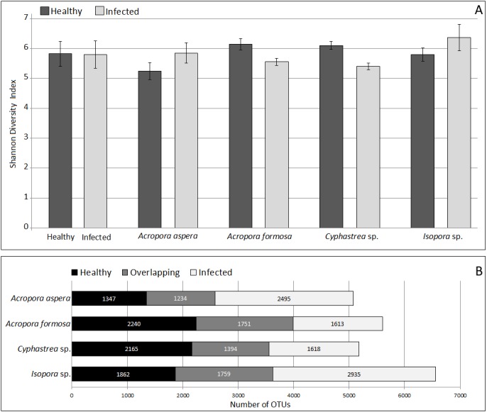
Coral reef ecosystems are impacted by climate change and human activities, such as increasing coastal development, overfishing, sewage and other pollutant discharge, and consequent eutrophication, which triggers increasing incidents of diseases and deterioration of corals worldwide. In this study, bacterial communities associated with four species of corals: Acropora aspera, Acropora formosa, Cyphastrea sp., and Isopora sp. in the healthy and disease stages with different diseases were compared using tagged 16S rRNA sequencing. In total, 59 bacterial phyla, 190 orders, and 307 genera were assigned in coral metagenomes where Proteobacteria and Firmicutes were pre-dominated followed by Bacteroidetes together with Actinobacteria, Fusobacteria, and Lentisphaerae as minor taxa. Principal Coordinates Analysis (PCoA) showed separated clustering of bacterial diversity in healthy and infected groups for individual coral species. Fusibacter was found as the major bacterial genus across all corals. The lower number of Fusibacter was found in A. aspera infected with white band disease and Isopora sp. with white plaque disease, but marked increases of Vibrio and Acrobacter, respectively, were observed. This was in contrast to A. formosa infected by a black band and Cyphastrea sp. infected by yellow blotch diseases which showed an increasing abundance of Fusibacter but a decrease in WH1-8 bacteria. Overall, infection was shown to result in disturbance in the complexity and structure of the associated bacterial microbiomes which can be relevant to the pathogenicity of the microbes associated with infected corals.
Keywords: 16S rRNA; Bacterial diversity; Coral; Metagenome; Next generation sequencing; Pathogenicity.
©2019 Mhuantong et al.
Conflict of interest statement
The authors declare there are no competing interests.
Figures


Similar articles
-
Identification of Candidate Coral Pathogens on White Band Disease-Infected Staghorn Coral.PLoS One. 2015 Aug 4;10(8):e0134416. doi: 10.1371/journal.pone.0134416. eCollection 2015. PLoS One. 2015. PMID: 26241853 Free PMC article.
-
The unexpected diversity of microbial communities associated with black corals revealed by high-throughput Illumina sequencing.FEMS Microbiol Lett. 2018 Aug 1;365(15). doi: 10.1093/femsle/fny167. FEMS Microbiol Lett. 2018. PMID: 29982506
-
Metagenomic data of bacterial communities associated with Acropora species from Phu Quoc Islands, Vietnam.Data Brief. 2023 Feb 14;47:108977. doi: 10.1016/j.dib.2023.108977. eCollection 2023 Apr. Data Brief. 2023. PMID: 36860407 Free PMC article.
-
Abundance and Multilocus Sequence Analysis of Vibrio Bacteria Associated with Diseased Elkhorn Coral (Acropora palmata) of the Florida Keys.Appl Environ Microbiol. 2018 Jan 2;84(2):e01035-17. doi: 10.1128/AEM.01035-17. Print 2018 Jan 15. Appl Environ Microbiol. 2018. PMID: 29079623 Free PMC article.
-
Complex interactions between potentially pathogenic, opportunistic, and resident bacteria emerge during infection on a reef-building coral.FEMS Microbiol Ecol. 2017 Jul 1;93(7). doi: 10.1093/femsec/fix080. FEMS Microbiol Ecol. 2017. PMID: 28637338
Cited by
-
Full-Length Transcriptome Maps of Reef-Building Coral Illuminate the Molecular Basis of Calcification, Symbiosis, and Circadian Genes.Int J Mol Sci. 2022 Sep 22;23(19):11135. doi: 10.3390/ijms231911135. Int J Mol Sci. 2022. PMID: 36232445 Free PMC article.
-
The Impact of Highly Weathered Oil from the Most Extensive Oil Spill in Tropical Oceans (Brazil) on the Microbiome of the Coral Mussismilia harttii.Microorganisms. 2023 Jul 29;11(8):1935. doi: 10.3390/microorganisms11081935. Microorganisms. 2023. PMID: 37630495 Free PMC article.
-
Identification of quorum sensing-regulated Vibrio fortis as potential pathogenic bacteria for coral bleaching and the effects on the microbial shift.Front Microbiol. 2023 Feb 3;14:1116737. doi: 10.3389/fmicb.2023.1116737. eCollection 2023. Front Microbiol. 2023. PMID: 36819038 Free PMC article.
-
Sedimentary Vibrio Blooms in the Xisha Islands May Associate with the 2020 Coral Bleaching Event.Appl Environ Microbiol. 2023 Jul 26;89(7):e0054323. doi: 10.1128/aem.00543-23. Epub 2023 Jun 14. Appl Environ Microbiol. 2023. PMID: 37314342 Free PMC article.
References
LinkOut - more resources
Full Text Sources
