. 2020 Feb 1;318(2):C422-C429.
doi: 10.1152/ajpcell.00430.2019. Epub 2019 Dec 25.

Rats genetically selected for low and high aerobic capacity exhibit altered soleus muscle myofilament functions

Affiliations

Rats genetically selected for low and high aerobic capacity exhibit altered soleus muscle myofilament functions

B J Biesiadecki et al. Am J Physiol Cell Physiol. .

Abstract

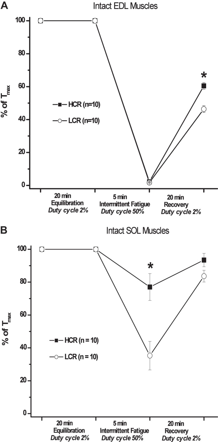
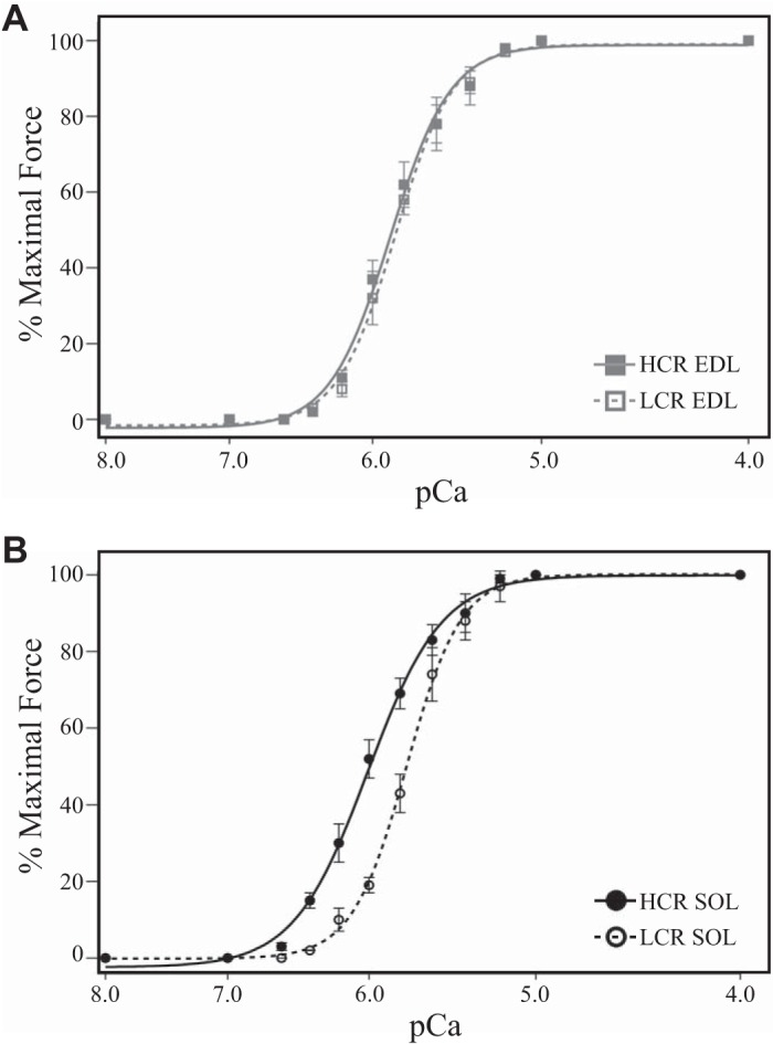
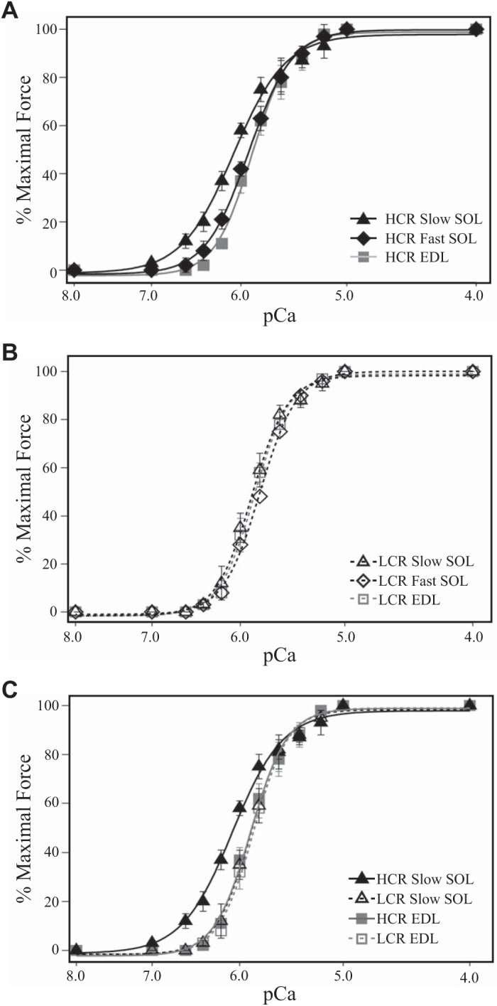
Aerobic exercise capacity is critical to bodily health. As a model to investigate the mechanisms that determine health and disease, we employed low (LCR) and high (HCR) capacity running rat models selectively bred to concentrate the genes responsible for divergent aerobic running capacity. To investigate the skeletal muscle contribution to this innate difference in running capacity we employed an approach combining examination of the myofilament protein composition and contractile properties of the fast fiber extensor digitorum longus (EDL) and slow fiber soleus (SOL) muscles from LCR and HCR rats. Intact muscle force experiments demonstrate that SOL, but not EDL, muscles from LCR rats exhibit a three times greater decrease in fatigued force. To investigate the mechanism of this increased fatigability in the LCR SOL muscle, we determined the myofilament protein composition and functional properties. Force-Ca2+ measurements demonstrate decreased Ca2+ sensitivity of single skinned SOL muscle fibers from LCR compared with that of HCR rats. Segregating SOL fibers into fast and slow types demonstrates that the decreased Ca2+ sensitivity in LCR SOL results from a specific decrease in slow-type SOL fiber Ca2+ sensitivity such that it was similar to that of fast-type fibers. These results identify that the altered myofilament contractile properties of LCR SOL slow-type fibers result in a fast muscle type Ca2+ sensitivity and the LCR muscle phenotype. Overall our findings demonstrate alterations of the myofilament proteins could contribute to fatigability of the SOL muscle and the decreased innate aerobic running performance of LCR compared with HCR rats.

Keywords: contractile proteins; muscle fatigue; myosin isoforms; single fiber; troponin isoforms.


Conflict of interest statement

No conflicts of interest, financial or otherwise, are declared by the authors.

Figures

Fig. 1.
Fig. 1.
Intact ex vivo muscle force development. Force development of isolated muscles was determined before, following 5-min intermittent fatigue and after 20-min recovery and represented as a percentage of maximal tetanic force at the end of the equilibration period (% of Tmax). A: force of isolated extensor digitorum longus (EDL) muscles was similarly decreased in high capacity runner (HCR; black square) and low capacity runner (LCR; white circle) muscles following fatigue treatment and LCR force remained decreased following recovery compared with HCR. B: force of isolated soleus (SOL) LCR muscle was half that of HCR following fatigue but similar after recovery. *P < 0.05.
Fig. 2.
Fig. 2.
Skinned fiber force-pCa relationship. A: the Ca2+-sensitive force development of high capacity runner (HCR; gray square) and low capacity runner (LCR; white square) extensor digitorum longus (EDL) fibers was minimally different. B: the Ca2+-sensitive force production of LCR soleus (SOL; white circle) fibers was significantly decreased compared with that of HCR SOL (black circle) fibers.
Fig. 3.
Fig. 3.
Soleus (SOL) fiber myofilament protein isoform expression. A: silver stain of representative single SOL fiber homogenates demonstrating two classes of SOL fibers that express either slow troponin (Tn)T, slow TnI, and slow myosin heavy chain (MHC)-I (Slow SOL) or fast TnT, fast TnI, and mixed slow MHC-I and fast MHC-IIa and MHC-IIx (Fast SOL). BD: Western blots of representative single Slow SOL and Fast SOL fiber homogenates with mAb CT3 (slow skeletal muscle TnT specific) alone or in combination with mAb T12 (fast skeletal muscle TnT specific) (B), mAb TnI-1 (fast and slow skeletal muscle TnI) (C), and silver stain or FA2 (type I slow MHC specific) (D). Mr Mkr, molecular marker.
Fig. 4.
Fig. 4.
Skinned force-pCa relationship in fast and slow-type muscle fibers. A: Ca2+-sensitive force development of high capacity runner (HCR) Slow soleus (SOL) fibers (black triangle) was increased compared with both HCR Fast SOL (black diamond) and extensor digitorum longus (EDL; gray square) fibers. B: contrary to its slow troponin isoform content, the Ca2+-sensitive force development of low capacity runner (LCR) Slow SOL (white triangle) fibers was not different from LCR Fast SOL (white diamond) or EDL (white square) fibers. C: Ca2+-sensitive force development of HCR Slow SOL fibers was increased compared with that of LCR Slow SOL that was not different from EDL fibers.

References

    1. Ackermann MA, Kerr JP, King B, Ward CW, Kontrogianni-Konstantopoulos A. The phosphorylation profile of myosin binding protein-C slow is dynamically regulated in slow-twitch muscles in health and disease. Sci Rep 5: 12637, 2015. [Erratum in Sci Rep 8: 46969, 2018]. doi:10.1038/srep12637. - DOI - PMC - PubMed
    1. Akar FG, Aon MA, Tomaselli GF, O’Rourke B. The mitochondrial origin of postischemic arrhythmias. J Clin Invest 115: 3527–3535, 2005. doi:10.1172/JCI25371. - DOI - PMC - PubMed
    1. Allen DG, Lamb GD, Westerblad H. Skeletal muscle fatigue: cellular mechanisms. Physiol Rev 88: 287–332, 2008. doi:10.1152/physrev.00015.2007. - DOI - PubMed
    1. Baldwin KM, Cooke DA, Cheadle WG. Time course adaptations in cardiac and skeletal muscle to different running programs. J Appl Physiol 42: 267–272, 1977. doi:10.1152/jappl.1977.42.2.267. - DOI - PubMed
    1. Brotto MA, Andreatta-van Leyen S, Nosek CM, Brotto LS, Nosek TM. Hypoxia and fatigue-induced modification of function and proteins in intact and skinned murine diaphragm muscle. Pflugers Arch 440: 727–734, 2000. doi:10.1007/s004240000327. - DOI - PubMed

Publication types