. 2020 Jan 1;161(1):bqz039.
doi: 10.1210/endocr/bqz039.

The Autophagy Gene Atg16L1 is Necessary for Endometrial Decidualization

Affiliations

The Autophagy Gene Atg16L1 is Necessary for Endometrial Decidualization

Arin K Oestreich et al. Endocrinology. .

Abstract

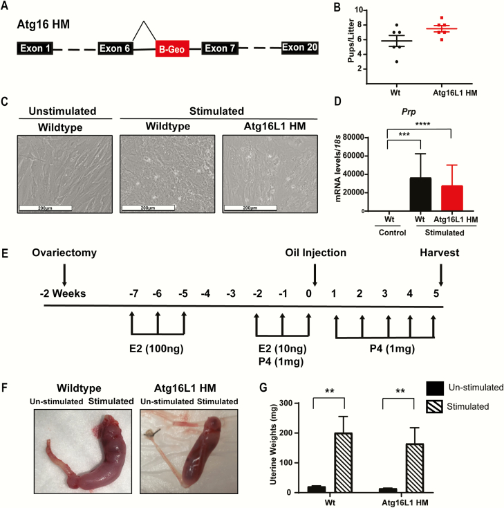
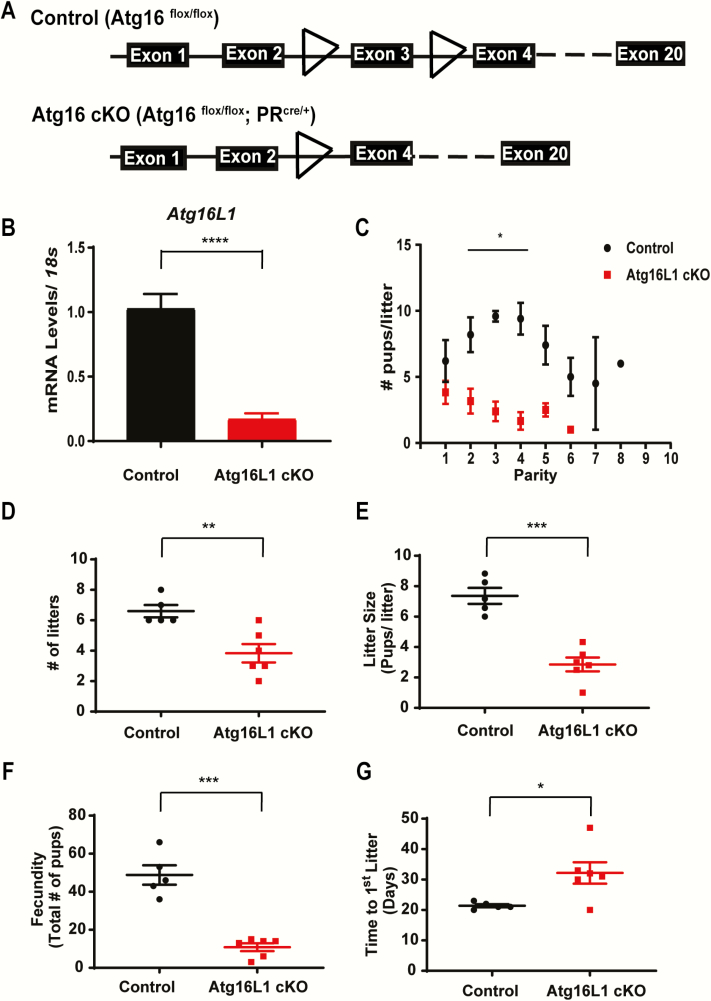
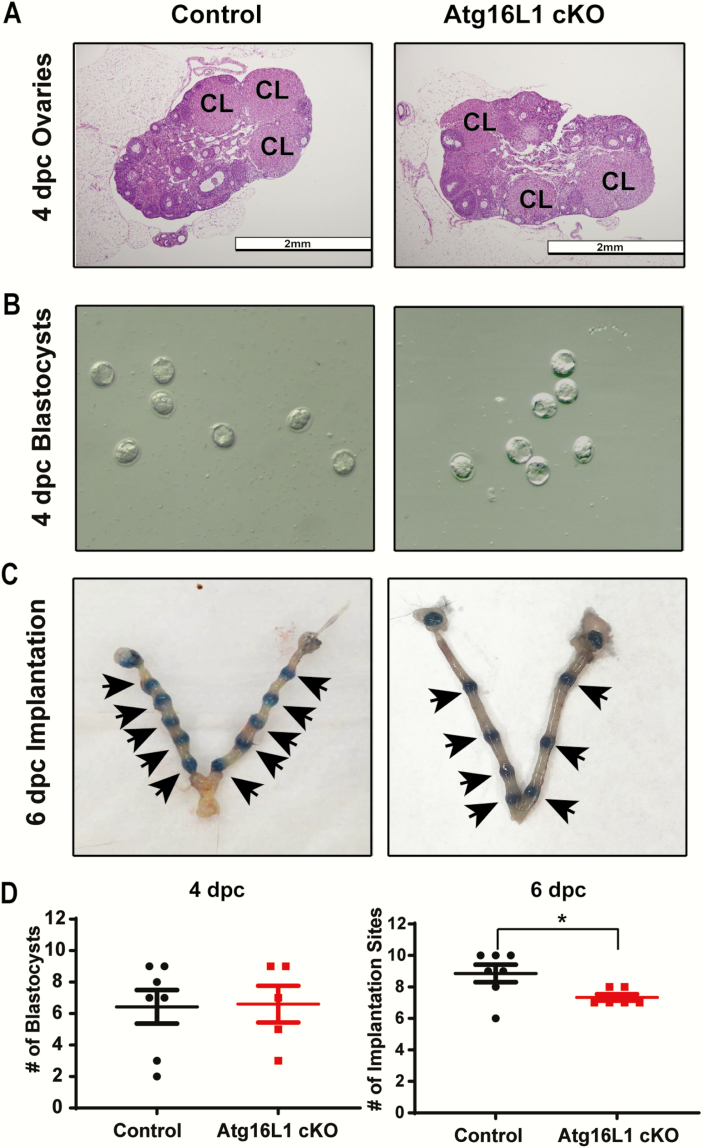
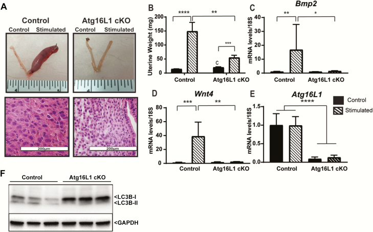
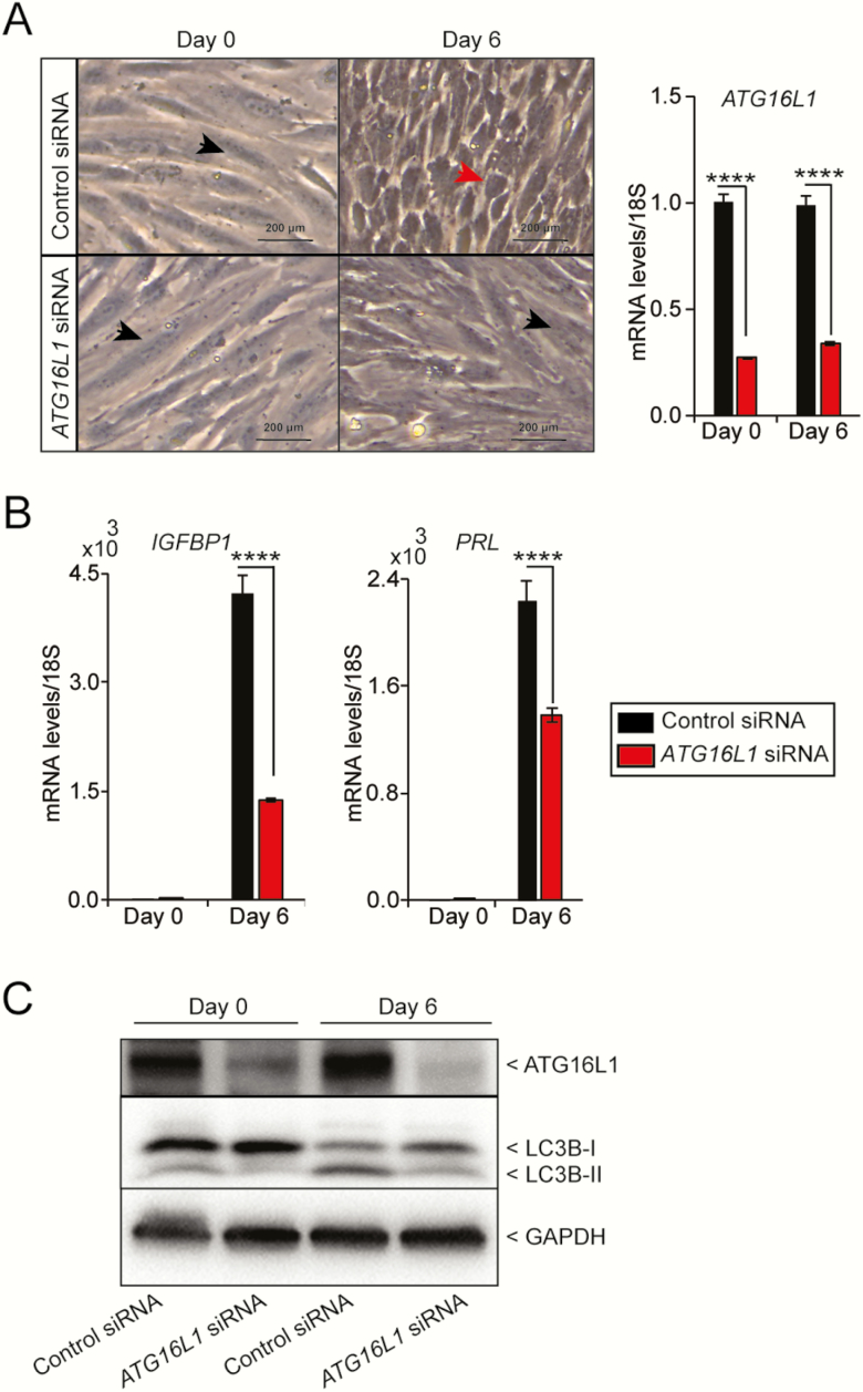
Uterine receptivity is critical for establishing and maintaining pregnancy. For the endometrium to become receptive, stromal cells must differentiate into decidual cells capable of secreting factors necessary for embryo survival and placental development. Although there are multiple reports of autophagy induction correlated with endometrial stromal cell (ESC) decidualization, the role of autophagy in decidualization has remained elusive. To determine the role of autophagy in decidualization, we utilized 2 genetic models carrying mutations to the autophagy gene Atg16L1. Although the hypomorphic Atg16L1 mouse was fertile and displayed proper decidualization, conditional knockout in the reproductive tract of female mice reduced fertility by decreasing the implantation rate. In the absence of Atg16L1, ESCs failed to properly decidualize and fewer blastocysts were able to implant. Additionally, small interfering RNA knock down of Atg16L1 was detrimental to the decidualization response of human ESCs. We conclude that Atg16L1 is necessary for decidualization, implantation, and overall fertility in mice. Furthermore, considering its requirement for human endometrial decidualization, these data suggest Atg16L1 may be a potential mediator of implantation success in women.

Keywords: fertility; implantation; pregnancy.


Figures

Figure 1.
Figure 1.
Atg16L1 HM mice are fertile and maintain the ability to decidualize. A) Diagram of the Atg16L1 HM mutation. B) Live birth rate of Wildtype (Wt) and Atg16L1 HM mice reveals that Atg16L1 HM mice are fertile. C) In vitro hormonal decidualization of endometrial stromal cells from Wt and Atg16L1 HM mice and D) Expression of the decidualization marker Prp show proper decidualization in Atg16L1 mice (n = 3 Wt, 5 Atg16L1 HM). E) Diagram of in vivo artificial decidualization method. F) Uterine horns of Wt and Atg16L1 HM mice and G) Resulting wet weights demonstrate Atg16L1 HM mice maintain the ability to decidualize in vivo (n = 10 Wt, 12 Atg16L1 HM). E2-estrogen, P4-progesterone; **P < 0.01, ***P < 0.001. ****P < 0.0001.
Figure 2.
Figure 2.
Uterine specific knock out of Atg16L1 impairs fertility. A) Diagram of the Atg16L1 cKO mutation. B) Expression of Atg16L1 is decreased in the uterus of Atg16L1 cKO virgin mice (n = 6 Control, 7 Atg16L1 cKO). C) Live birth rate, D) Number of litters, E) Litter size, and F) Total number of pups produced by control and Atg16L1 cKO mice over a 6-month period reveals compromised fertility in Atg16L1 mice (n = 5 Controls, 6 Atg16L1 cKO). G) Time to first litter as measured by plug date to delivery date is extended in Atg16L1 cKO mice; *P < 0.05, **P < 0.01, ***P < 0.001. ****P < 0.0001.
Figure 3.
Figure 3.
Atg16L1 cKO mice have an implantation defect. A) Histological sections of 4 dpc ovaries from control and Atg16L1 cKO mice show normal morphology with the presence of corpora lutea (denoted CL) (n = 6 Control, 5 Atg16L1 cKO). B) Blastocysts flushed from the uterine horns of control and Atg16L1 cKO mice on 4 dpc show normal morphology (n = 7 Control, 5 Atg15L1 cKO pregnant dams). C) Implantation cites observed at 6 dpc in control and Atg16L1 cKO mice (n = 7 Control and 6 Atg16L1 cKO pregnant dams). D) Quantification of blastocysts at 4 dpc and implantation cites at 6 dpc reveal 2 fewer blastocysts implant in the Atg16L1 cKO uterus. Arrows indicate implantation sites; *P < 0.05
Figure 4.
Figure 4.
Atg16L1 mice have impaired decidualization. A) Dissected uteri, H&E histology, and B) Wet weights of control and Atg16L1 cKO mice that have been stimulated to decidualize (n = 13 Control and n = 11 Atg16L1 cKO). Expression of the decidualization markers C) Bmp2 and D) Wnt4 reveal impaired decidualization in Atg16L1 cKO mice. E) Expression of Atg16L1 in control and Atg16L1 cKO mice following artificial decidualization, (n = 6 Control, 5 Atg16L1 cKO). F) Immunoblotting of LC3B protein on the protein lysate collected from stimulated horn of Control and Atg16L1 cKO mice uteri (n = 3) mice. The GAPDH is used as an internal loading control; *P < 0.05, **P < 0.01, ***P < 0.001. ****P < 0.0001.
Figure 5.
Figure 5.
Knock down of ATG16L1 in human endometrial stromal cells impairs decidualization. A) Cellular morphology and B) quantitative PCR analysis of the decidualization marker IGFBP1 and PRL following transfection with control or ATG16L1 siRNA and hormonal stimulation. Data is normalized to 18S and expressed as fold change over Day 0 controls. Results are shown as mean ± standard error (SE) from 3 replicates of 1 patient-derived primary endometrial cell line. The experiment carried out on hESCs derived from 4 different individuals. (n = 4). Black arrowhead indicates fibroblast morphology and red arrowhead indicates decidualizing morphology. C) Immunoblotting of ATG16L1 and LC3B on the protein lysate from the hESCs collected at day 0 and day 6 after transfected with control or ATG16L1 siRNA. The GAPDH is used as an internal loading control; ****P < 0.0001.

References

    1. Paria BC, Wang H, Dey SK. Endocannabinoid signaling in synchronizing embryo development and uterine receptivity for implantation. Chem Phys Lipids. 2002;121(1-2): 201–210. - PubMed
    1. Psychoyos A. Uterine receptivity for nidation. Ann N Y Acad Sci. 1986;476:36–42. - PubMed
    1. Schlafke S, Enders AC. Cellular basis of interaction between trophoblast and uterus at implantation. Biol Reprod. 1975;12(1):41–65. - PubMed
    1. Lessey BA. Endometrial receptivity and the window of implantation. Baillieres Best Pract Res Clin Obstet Gynaecol. 2000;14(5):775–788. - PubMed
    1. Ola B, Li TC. Implantation failure following in-vitro fertilization. Curr Opin Obstet Gynecol. 2006;18(4):440–445. - PubMed

MeSH terms

Substances