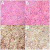
Solitary Fibrous Tumor of the Orbit: A Case Series With Clinicopathologic Correlation and Evaluation of STAT6 as a Diagnostic Marker
- PMID: 31876648
- PMCID: PMC7060103
- DOI: 10.1097/IOP.0000000000001504
Solitary Fibrous Tumor of the Orbit: A Case Series With Clinicopathologic Correlation and Evaluation of STAT6 as a Diagnostic Marker
Abstract
Purpose: To retrospectively describe the clinical characteristics, management, and outcomes of a series of patients with solitary fibrous tumor (SFT) of the orbit and to evaluate signal transducer and activator of transcription 6 (STAT6) as a diagnostic marker.
Methods: Review of a retrospective, noncomparative, consecutive series of patients treated at a single institution with a histopathologic diagnosis of SFT. Demographic, clinical, and imaging data were collected, and paraffin-embedded tissue sections were stained to evaluate for the presence of STAT6 and other pertinent markers.
Results: Twenty-one patients were identified. Most presented with painless progressive proptosis or eyelid swelling for less than 6 months. Imaging revealed well-circumscribed, firm, variably vascular contrast-enhancing lesions with low to medium reflectivity on ultrasound. Four tumors were histopathologically malignant. All tumors were primarily excised, and 1 patient required exenteration. Two patients were treated with adjuvant radiation therapy. Six patients had recurrent disease of which 3 underwent repeat excision, and 2 were observed. No metastatic disease or attributable deaths were observed. All lesions with available tissue stained positively for both CD34 and STAT6.
Conclusion: This is the largest single institution case series of orbital SFT with clinicopathologic correlation and the largest series to confirm the presence of STAT6 in orbital lesions. The management of SFT remains challenging due to unpredictable tumor behavior, and complete excision is the generally recommended treatment. It remains unclear whether a subset of asymptomatic patients with histopathologically benign disease can be durably observed without negative sequelae.
Conflict of interest statement
Figures


Similar articles
-
Solitary Fibrous Tumors in Pediatric Patients: A Rare and Potentially Overdiagnosed Neoplasm, Confirmed by STAT6 Immunohistochemistry.Pediatr Dev Pathol. 2018 Jul-Aug;21(4):389-400. doi: 10.1177/1093526617745431. Epub 2017 Dec 11. Pediatr Dev Pathol. 2018. PMID: 29228868
-
[Expression and significance of STAT6 in solitary fibrous tumor].Zhonghua Bing Li Xue Za Zhi. 2017 Apr 8;46(4):235-239. doi: 10.3760/cma.j.issn.0529-5807.2017.04.004. Zhonghua Bing Li Xue Za Zhi. 2017. PMID: 28376588 Chinese.
-
Solitary fibrous tumor of the breast: report of a case with emphasis on diagnostic role of STAT6 immunostaining.Pathol Res Pract. 2016 May;212(5):463-7. doi: 10.1016/j.prp.2015.12.013. Epub 2015 Dec 31. Pathol Res Pract. 2016. PMID: 26778386
-
Ocular adnexal (orbital) solitary fibrous tumor: nuclear STAT6 expression and literature review.Graefes Arch Clin Exp Ophthalmol. 2015 Sep;253(9):1609-17. doi: 10.1007/s00417-015-2975-5. Epub 2015 Mar 13. Graefes Arch Clin Exp Ophthalmol. 2015. PMID: 25761539 Review.
-
Meningeal solitary fibrous tumor/hemangiopericytoma: Emphasizing on STAT 6 immunohistochemistry with a review of literature.Neurol India. 2018 Sep-Oct;66(5):1419-1426. doi: 10.4103/0028-3886.241365. Neurol India. 2018. PMID: 30233017 Review.
Cited by
-
Presentation of orbital solitary fibrous tumours.Eye (Lond). 2023 Nov;37(16):3406-3411. doi: 10.1038/s41433-023-02519-7. Epub 2023 Apr 15. Eye (Lond). 2023. PMID: 37061621 Free PMC article.
-
Orbital Solitary Fibrous Tumor: Four Case Reports-Clinical and Histopathological Features.Case Rep Ophthalmol Med. 2021 Jun 9;2021:5822859. doi: 10.1155/2021/5822859. eCollection 2021. Case Rep Ophthalmol Med. 2021. PMID: 34211794 Free PMC article.
-
A challenging case of solitary fibrous tumor of the orbit in an anemic patient.Rom J Ophthalmol. 2024 Oct-Dec;68(4):457-461. doi: 10.22336/rjo.2024.82. Rom J Ophthalmol. 2024. PMID: 39936050 Free PMC article.
-
Rapidly growing solitary fibrous tumors of the pleura: a case report and review of the literature.Ann Transl Med. 2020 Jul;8(14):890. doi: 10.21037/atm-20-4974. Ann Transl Med. 2020. PMID: 32793734 Free PMC article.
-
Recurrent solitary fibrous tumor of eyelid: A rare entity.Oman J Ophthalmol. 2023 Feb 21;16(1):103-105. doi: 10.4103/ojo.ojo_135_22. eCollection 2023 Jan-Apr. Oman J Ophthalmol. 2023. PMID: 37007226 Free PMC article.
References
-
- Fletcher CDM, World Health Organization., International Agency for Research on Cancer. WHO classification of tumours of soft tissue and bone, 4th ed. Lyon: IARC Press, 2013; 468 p.
-
- Westra WH, Gerald WL, Rosai J. Solitary fibrous tumor. Consistent CD34 immunoreactivity and occurrence in the orbit. Am J Surg Pathol 1994;18(10):992–8. - PubMed
-
- Alexandrakis G, Johnson TE. Recurrent orbital solitary fibrous tumor in a 14-year-old girl. Am J Ophthalmol 2000;130(3):373–6. - PubMed
Publication types
MeSH terms
Substances
Grants and funding
LinkOut - more resources
Full Text Sources
Research Materials
Miscellaneous

