

Confocal Microscopy: Principles and Modern Practices
- PMID: 31876974
- PMCID: PMC6961134
- DOI: 10.1002/cpcy.68
Confocal Microscopy: Principles and Modern Practices
Abstract
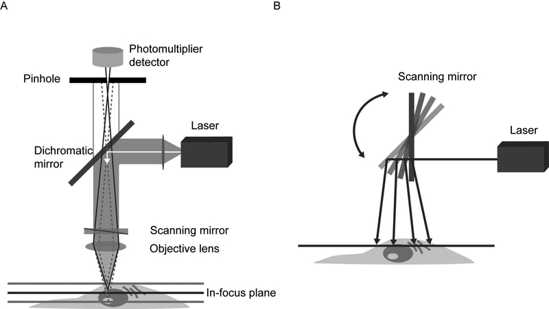
In light microscopy, illuminating light is passed through the sample as uniformly as possible over the field of view. For thicker samples, where the objective lens does not have sufficient depth of focus, light from sample planes above and below the focal plane will also be detected. The out-of-focus light will add blur to the image, reducing the resolution. In fluorescence microscopy, any dye molecules in the field of view will be stimulated, including those in out-of-focus planes. Confocal microscopy provides a means of rejecting the out-of-focus light from the detector such that it does not contribute blur to the images being collected. This technique allows for high-resolution imaging in thick tissues. In a confocal microscope, the illumination and detection optics are focused on the same diffraction-limited spot in the sample, which is the only spot imaged by the detector during a confocal scan. To generate a complete image, the spot must be moved over the sample and data collected point by point. A significant advantage of the confocal microscope is the optical sectioning provided, which allows for 3D reconstruction of a sample from high-resolution stacks of images. Several types of confocal microscopes have been developed for this purpose, and each has different advantages and disadvantages. This article provides a concise introduction to confocal microscopy. © 2019 by John Wiley & Sons, Inc.
Keywords: confocal microscopy; fluorescence; laser scanning; resonant scanning; spinning disk.
© 2019 John Wiley & Sons, Inc.
Figures

References
-
- Bolbat A, & Schultz C 2016. Recent developments of genetically encoded optical sensors for cell biology. Molecular Biology of the Cell. - PubMed
-
- Brown CM 2007. Fluorescence microscopy – avoiding the pitfalls. Journal of Cell Science. 120:1703–1705. - PubMed
-
- Callamaras N & Parker I 1999. Construction of a confocal microscope for real-time x-y and x-z imaging. Cell Calcium, 26(6):271–279. - PubMed
Publication types
MeSH terms
Grants and funding
LinkOut - more resources
Full Text Sources
Other Literature Sources
