

Clinical and Genetic Analysis of a European Cohort with Pericentral Retinitis Pigmentosa
- PMID: 31877679
- PMCID: PMC6982348
- DOI: 10.3390/ijms21010086
Clinical and Genetic Analysis of a European Cohort with Pericentral Retinitis Pigmentosa
Abstract
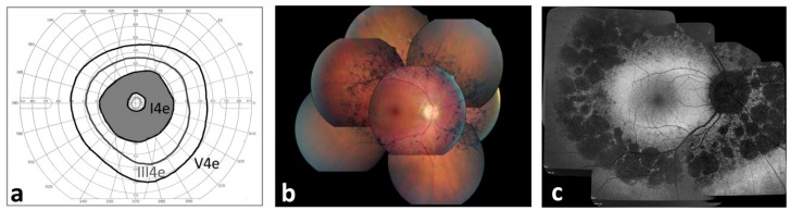
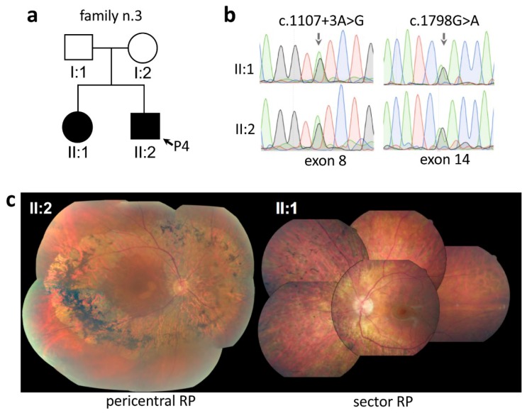
Retinitis pigmentosa (RP) is a clinically heterogenous disease that comprises a wide range of phenotypic and genetic subtypes. Pericentral RP is an atypical form of RP characterized by bone-spicule pigmentation and/or atrophy confined in the near mid-periphery of the retina. In contrast to classic RP, the far periphery is better preserved in pericentral RP. The aim of this study was to perform the first detailed clinical and genetic analysis of a cohort of European subjects with pericentral RP to determine the phenotypic features and the genetic bases of the disease. A total of 54 subjects from 48 independent families with pericentral RP, non-syndromic and syndromic, were evaluated through a full ophthalmological examination and underwent clinical exome or retinopathy gene panel sequencing. Disease-causative variants were identified in 22 of the 35 families (63%) in 10 different genes, four of which are also responsible for syndromic RP. Thirteen of the 34 likely pathogenic variants were novel. Intra-familiar variability was also observed. The current study confirms the mild phenotype of pericentral RP and extends the spectrum of genes associated with this condition.
Keywords: USH2A; inherited retinal dystrophies; next generation sequencing; pericentral retinitis pigmentosa.
Conflict of interest statement
The authors declare no conflict of interest.
Figures

References
-
- Matsui R., Cideciyan A.V., Schwartz S.B., Sumaroka A., Roman A.J., Swider M., Huang W.C., Sheplock R., Jacobson S.G. Molecular heterogeneity within the clinical diagnosis of pericentral retinal degeneration. Investig. Ophthalmol. Vis. Sci. 2015;56:6007–6018. doi: 10.1167/iovs.15-17174. - DOI - PMC - PubMed
MeSH terms
Supplementary concepts
Grants and funding
LinkOut - more resources
Full Text Sources
