

Nanomaterials for Biosensing Lipopolysaccharide
- PMID: 31877825
- PMCID: PMC7168309
- DOI: 10.3390/bios10010002
Nanomaterials for Biosensing Lipopolysaccharide
Abstract
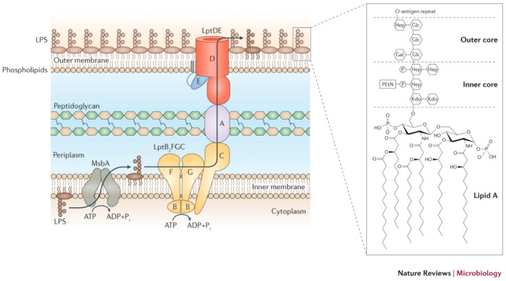
Lipopolysaccharides (LPS) are endotoxins, hazardous and toxic inflammatory stimulators released from the outer membrane of Gram-negative bacteria, and are the major cause of septic shock giving rise to millions of fatal illnesses worldwide. There is an urgent need to identify and detect these molecules selectively and rapidly. Pathogen detection has been done by traditional as well as biosensor-based methods. Nanomaterial based biosensors can assist in achieving these goals and have tremendous potential. The biosensing techniques developed are low-cost, easy to operate, and give a fast response. Due to extremely small size, large surface area, and scope for surface modification, nanomaterials have been used to target various biomolecules, including LPS. The sensing mechanism can be quite complex and involves the transformation of chemical interactions into amplified physical signals. Many different sorts of nanomaterials such as metal nanomaterials, magnetic nanomaterials, quantum dots, and others have been used for biosensing of LPS and have shown attractive results. This review considers the recent developments in the application of nanomaterials in sensing of LPS with emphasis given mainly to electrochemical and optical sensing.
Keywords: biosensing; endotoxin; lipopolysaccharides (LPS); nanomaterials.
Conflict of interest statement
The authors declare no conflict of interest.
Figures
Similar articles
-
High-Performance Biosensing Systems Based on Various Nanomaterials as Signal Transducers.Biotechnol J. 2019 Jan;14(1):e1800249. doi: 10.1002/biot.201800249. Epub 2018 Aug 28. Biotechnol J. 2019. PMID: 30117715 Review.
-
Nanomaterials-based enzyme electrochemical biosensors operating through inhibition for biosensing applications.Biosens Bioelectron. 2017 Mar 15;89(Pt 2):886-898. doi: 10.1016/j.bios.2016.09.102. Epub 2016 Sep 29. Biosens Bioelectron. 2017. PMID: 27818056 Review.
-
Recent advances in nanomaterial-based electrochemical and optical sensing platforms for microRNA assays.Analyst. 2019 May 7;144(9):2849-2866. doi: 10.1039/c9an00081j. Epub 2019 Mar 27. Analyst. 2019. PMID: 30916675 Review.
-
Advances in endotoxin analysis.Adv Clin Chem. 2024;118:1-34. doi: 10.1016/bs.acc.2023.11.001. Epub 2024 Jan 15. Adv Clin Chem. 2024. PMID: 38280803
-
Metal oxide nanomaterials based electrochemical and optical biosensors for biomedical applications: Recent advances and future prospectives.Environ Res. 2024 Apr 15;247:118002. doi: 10.1016/j.envres.2023.118002. Epub 2023 Dec 25. Environ Res. 2024. PMID: 38151147 Review.
Cited by
-
α-Tocopherol Protects Lipopolysaccharide-Activated BV2 Microglia.Molecules. 2023 Apr 10;28(8):3340. doi: 10.3390/molecules28083340. Molecules. 2023. PMID: 37110573 Free PMC article.
-
Wettability-based ultrasensitive detection of amphiphiles through directed concentration at disordered regions in self-assembled monolayers.Proc Natl Acad Sci U S A. 2022 Oct 25;119(43):e2211042119. doi: 10.1073/pnas.2211042119. Epub 2022 Oct 17. Proc Natl Acad Sci U S A. 2022. PMID: 36252006 Free PMC article.
-
A Review on Low-Dimensional Nanomaterials: Nanofabrication, Characterization and Applications.Nanomaterials (Basel). 2022 Dec 29;13(1):160. doi: 10.3390/nano13010160. Nanomaterials (Basel). 2022. PMID: 36616070 Free PMC article. Review.
-
Avian pathogenic Escherichia coli: Epidemiology, virulence and pathogenesis, diagnosis, pathophysiology, transmission, vaccination, and control.Vet World. 2024 Dec;17(12):2747-2762. doi: 10.14202/vetworld.2024.2747-2762. Epub 2024 Dec 6. Vet World. 2024. PMID: 39897356 Free PMC article. Review.
-
Application of Nanomaterial-Mediated Ferroptosis Regulation in Kidney Disease.Int J Nanomedicine. 2025 Feb 5;20:1637-1659. doi: 10.2147/IJN.S496644. eCollection 2025. Int J Nanomedicine. 2025. PMID: 39931533 Free PMC article. Review.
References
Publication types
MeSH terms
Substances
LinkOut - more resources
Full Text Sources
Molecular Biology Databases
