Review
. 2019 Dec 22;21(1):106.
doi: 10.3390/ijms21010106.

Effects of Gangliosides on Spermatozoa, Oocytes, and Preimplantation Embryos

Affiliations
Review

Effects of Gangliosides on Spermatozoa, Oocytes, and Preimplantation Embryos

Bo Hyun Kim et al. Int J Mol Sci. .

Abstract

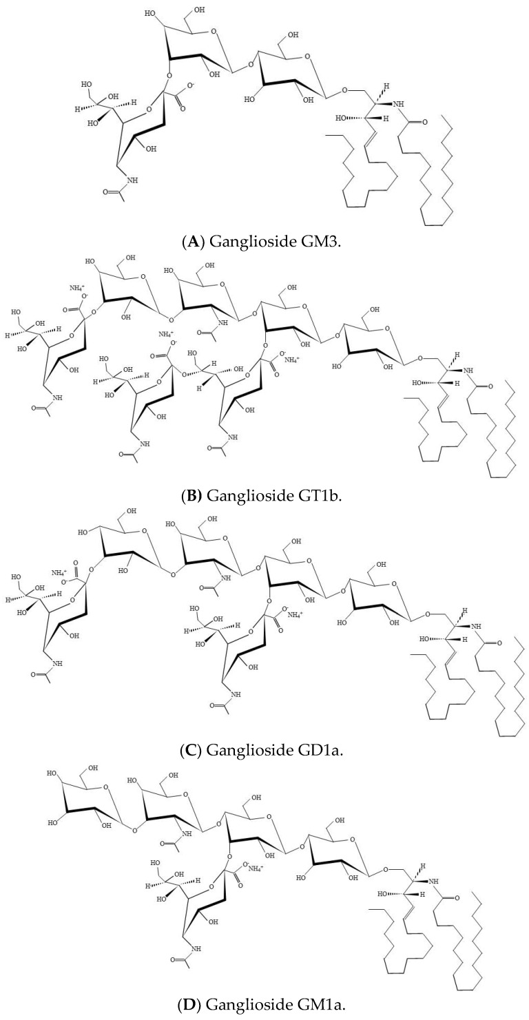
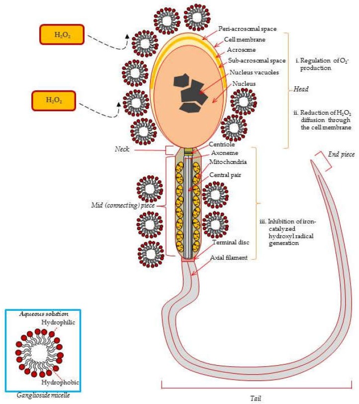
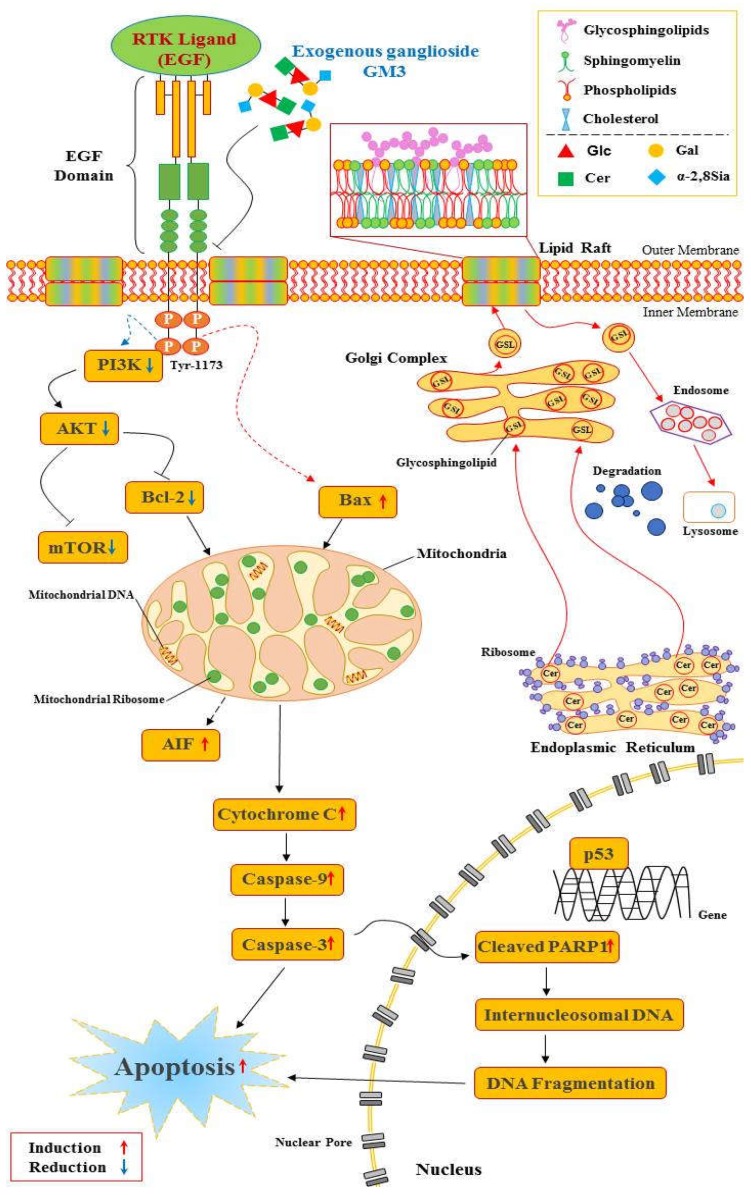
Gangliosides are sialic acid-containing glycosphingolipids, which are the most abundant family of glycolipids in eukaryotes. Gangliosides have been suggested to be important lipid molecules required for the control of cellular procedures, such as cell differentiation, proliferation, and signaling. GD1a is expressed in interstitial cells during ovarian maturation in mice and exogenous GD1a is important to oocyte maturation, monospermic fertilization, and embryonic development. In this context, GM1 is known to influence signaling pathways in cells and is important in sperm-oocyte interactions and sperm maturation processes, such as capacitation. GM3 is expressed in the vertebrate oocyte cytoplasm, and exogenously added GM3 induces apoptosis and DNA injury during in vitro oocyte maturation and embryogenesis. As a consequence of this, ganglioside GT1b and GM1 decrease DNA fragmentation and act as H2O2 inhibitors on germ cells and preimplantation embryos. This review describes the functional roles of gangliosides in spermatozoa, oocytes, and early embryonic development.

Keywords: apoptosis; embryo; ganglioside; maturation; oocyte; spermatozoa.


Conflict of interest statement

The authors declare no conflicts of interest.

Figures

Figure 1
Figure 1
Ganglioside structure. (A) Structure of GM3 monoganglioside. (B) Structure of trisialoganglioside GT1b. (C) Structure of GD1a. (D) Structure of GM1a.
Figure 2
Figure 2
Protective effects of exogenous ganglioside micelles from ROS-induced damage in the ejaculated spermatozoa. Exogenous gangliosides are absorbed to the sperm surface and reduce the superoxide anion level generated by PMA-stimulated spermatozoa. O-2 is converted into H2O2 by cytosolic-localized SOD1 (Cu, Zn, SOD), mitochondria-localized SOD2 (Mn SOD), and the extracellular SOD3 (Fe SOD). Ganglioside GT1b reduces Fe2+-mediated decomposition of lipid hydroperoxydes from the sperm membrane. Consequently, GT1b prevents lipid peroxidation induced sperm membrane damage due to its specific molecular structure, such as micelles [16,20,70].
Figure 3
Figure 3
Exogenous GM3 and the EGFR-mediated PI3K/AKT signal pathway. Exogenously added GM3 binds to the extracellular domain of EGFR and inhibits its dimerization [104] without inhibiting ligand binding [105]. Exogenous GM3 inhibits EGF-induced phosphorylation of EGFR at the Tyr-1173 residue [35]. After inhibition by blocking phosphorylation, the inhibited signal from the EGFR prevents one of the three downstream signaling pathways, PI3K, AKT, or mTOR. PI3K and AKT activation is reduced [106]. Upon inhibition of the phosphorylation of PI3K, AKT subsequently suppresses the phosphorylation of mTOR, which mediates protein synthesis [107]. This mechanism for suppressed EGFR activity might reduce cell proliferation and inhibit the repair of DNA damage [108]. The activation changes of these pro- and anti-apoptotic proteins is initiated in mitochondria [109]. Continuous mitochondrial regulation releases pro-apoptotic proteins such as cytochrome c into the cytoplasm causing caspase cascades processed by apoptosis, the programed cell death [52].The most compelling evidence shows that GM3 treatment decreases phosphorylated EGFR levels and regulates down-stream EGFR activation in cumulus-oocyte complexes COCs. Consequently, GM3 exposure reduces EGFR-delivered PI3K/AKT signaling for proliferation during in vitro maturation of porcine COCs [36].

Similar articles

Cited by

References

    1. Edidin M. The state of lipid rafts: From model membranes to cell. Ann. Rev. Biophys. Struct. 2003;32:257–283. doi: 10.1146/annurev.biophys.32.110601.142439. - DOI - PubMed
    1. Brown D. Lipid rafts, detergent-resistant membranes, and raft targeting signals. Physiology. 2006;21:430–439. doi: 10.1152/physiol.00032.2006. - DOI - PubMed
    1. Mclntosh T.J. Overview of membrane rafts. Meth. Mol. Biol. 2007;398:1–9. - PubMed
    1. Simons K., Ikonen E. Functional rafts in cell membranes. Nature. 1997;387:569–572. doi: 10.1038/42408. - DOI - PubMed
    1. Silvius J.R. Partitioning of membrane molecules between raft and non-raft domains: Insights from membrane studies. Biochem. Biophys. Acta. 2005;1746:193–202. doi: 10.1016/j.bbamcr.2005.09.003. - DOI - PubMed

Substances