. 2019 Dec 23;11(1):18.
doi: 10.3390/genes11010018.

Vulpinic Acid Controls Stem Cell Fate toward Osteogenesis and Adipogenesis

Affiliations

Vulpinic Acid Controls Stem Cell Fate toward Osteogenesis and Adipogenesis

Sang Ah Yi et al. Genes (Basel). .

Abstract

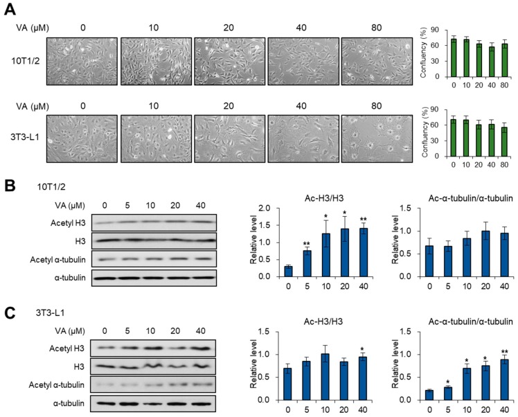
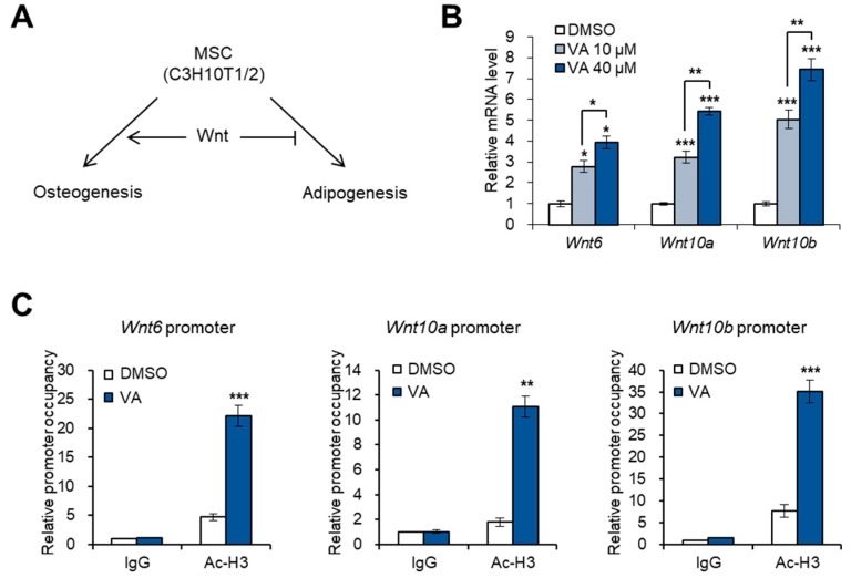
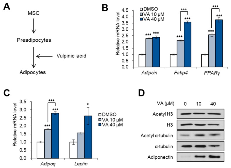
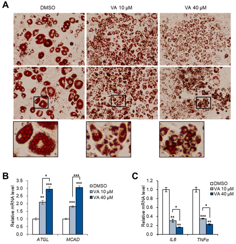
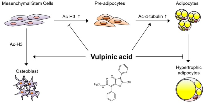
Vulpinic acid, a naturally occurring methyl ester of pulvinic acid, has been reported to exert anti-fungal, anti-cancer, and anti-oxidative effects. However, its metabolic action has not been implicated yet. Here, we show that vulpinic acid derived from a mushroom, Pulveroboletus ravenelii controls the cell fate of mesenchymal stem cells and preadipocytes by inducing the acetylation of histone H3 and α-tubulin, respectively. The treatment of 10T1/2 mesenchymal stem cells with vulpinic acid increased the expression of Wnt6, Wnt10a, and Wnt10b, which led to osteogenesis inhibiting the adipogenic lineage commitment, through the upregulation of H3 acetylation. By contrast, treatment with vulpinic acid promoted the terminal differentiation of 3T3-L1 preadipocytes into mature adipocytes. In this process, the increase in acetylated tubulin was accompanied, while acetylated H3 was not altered. As excessive generation of adipocytes occurs, the accumulation of lipid drops was not concentrated, but dispersed into a number of adipocytes. Consistently, the expressions of lipolytic genes were upregulated and inflammatory factors were downregulated in adipocytes exposed to vulpinic acid during adipogenesis. These findings reveal the multiple actions of vulpinic acid in two stages of differentiation, promoting the osteogenesis of mesenchymal stem cells and decreasing hypertrophic adipocytes, which can provide experimental evidence for the novel metabolic advantages of vulpinic acid.

Keywords: acetyl H3; acetyl tubulin; adipogenesis; cell fate; hypertrophy; osteogenesis; vulpinic acid.


Conflict of interest statement

The authors declare no conflict of interest.

Figures

Figure 1
Figure 1
Vulpinic acid (VA) modulates acetylation of histone H3 and α-tubulin in 10T1/2 and 3T3-L1 cells. (A) Microscopic images (100X) of 10T1/2 and 3T3-L1 cells treated with vulpinic acid for 24 h at the indicated concentrations. (B) Immunoblot analysis of 10T1/2 cells treated with vulpinic acid for 24 h at the indicated concentrations. (C) Immunoblot analysis of 3T3-L1 cells treated with vulpinic acid for 24 h at the indicated concentrations. Data represent means ± SEM (standard effort of the mean) for n = 3. * p < 0.05, ** p < 0.01.
Figure 2
Figure 2
Vulpinic acid promotes the expression of Wnt genes via H3 acetylation in 10T1/2 MSCs. (A) Schematic representation of Wnt signaling-mediated cell fate determination of mesenchymal stem cells (MSCs). (B) The mRNA levels of Wnt6, Wnt10a, and Wnt10b genes in 10T1/2 cells treated with vulpinic acid (10 or 40 μM) for 24 h. (C) 10T1/2 cells were treated with or without vulpinic acid (40 μM) for 24 h. ChIP assay was performed with IgG and acetylated H3 antibodies followed by real time PCR with primers for promoter regions of Wnt6, Wnt10a, and Wnt10b genes. Data represent means ± SEM for n = 3. * p < 0.05, ** p < 0.01, *** p < 0.001.
Figure 3
Figure 3
Vulpinic acid induces osteogenesis via H3 acetylation in 10T1/2 MSCs. (A) Schematic representation of BMPs-mediated osteogenic or adipogenic commitment of MSCs. (B) Immunoblot analysis of 10T1/2 cells treated with vulpinic acid (10 or 40 μM) for 24 h in the presence of BMP2 or BMP4. (C) 10T1/2 cells were treated with or without vulpinic acid (40 μM) for 24 h in the presence of BMP2 or BMP4. ChIP assay was performed with IgG and acetylated H3 antibodies followed by real time PCR with primers for promoter region of Runx2 gene. (D) The mRNA levels of the Bmp2, Ocn, and Sp7 genes in 10T1/2 cells treated with vulpinic acid (10 or 40 μM) for 24 h. (E) The mRNA levels of Cebpa and PPARγ genes in 10T1/2 cells treated with vulpinic acid (10 or 40 μM) for 24 h in the presence of BMP4. Data represent means ± SEM for n = 3. * p < 0.05, ** p < 0.01, *** p < 0.001.
Figure 4
Figure 4
Vulpinic acid promotes adipogenesis from 3T3-L1 preadipocytes. (A) Schematic representation of adipogenesis process. 3T3-L1 cells were incubated with an adipogenic medium in the absence or presence of vulpinic acid. (B) The mRNA levels of Adipsin, Fabp4, and PPARγ genes in 3T3-L1 adipocytes incubated with vulpinic acid (10 or 40 μM) during adipogenesis. (C) The mRNA levels of Adipoq and Leptin genes in 3T3-L1 adipocytes incubated with vulpinic acid (10 or 40 μM) during adipogenesis. (D) Immunoblot analysis of 3T3-L1 adipocytes incubated with vulpinic acid (10 or 40 μM) during adipogenesis. Data represent means ± SEM for n = 3. * p < 0.05, ** p < 0.01, *** p < 0.001.
Figure 5
Figure 5
Vulpinic acid reduces hypertrophic adipocytes by promoting hyperplasia. (A) Oil-red-O staining of 3T3-L1 adipocytes incubated with vulpinic acid (10 or 40 μM) during adipogenesis. Scale bar = 200 μm (40X). (B) The mRNA levels of ATGL and MCAD genes in 3T3-L1 adipocytes incubated with vulpinic acid (10 or 40 μM) during adipogenesis. (C) The mRNA levels of IL6 and TNFα genes in 3T3-L1 adipocytes incubated with vulpinic acid (10 or 40 μM) during adipogenesis. Data represent means ± SEM for n = 3. * p < 0.05, ** p < 0.01, *** p < 0.001.
Figure 6
Figure 6
Molecular model underlying the mechanism of actions of vulpinic acid. During the early stage of mesenchymal stem cells, vulpinic acid promotes osteogenic commitment while preventing adipogenic commitment via H3 acetylation-mediated gene modulations. During terminal differentiation to mature adipocytes from preadipocytes, vulpinic acid enhances de novo generation of adipocytes via acetylation of α-tubulin, reducing hypertrophic adipocytes.

References

    1. Baek S.C., Choi E., Eom H.J., Jo M.S., Kim S., So H.M., Kim S.H., Kang K.S., Kim K.H. LC/MS-based analysis of bioactive compounds from the bark of Betula platyphylla var. japonica and their effects on regulation of adipocyte and osteoblast differentiation. Nat. Prod. Sci. 2018;24:235–240. doi: 10.20307/nps.2018.24.4.235. - DOI
    1. Augello A., De Bari C. The regulation of differentiation in mesenchymal stem cells. Hum. Gene Ther. 2010;21:1226–1238. doi: 10.1089/hum.2010.173. - DOI - PubMed
    1. Chen Q., Shou P., Zheng C., Jiang M., Cao G., Yang Q., Cao J., Xie N., Velletri T., Zhang X., et al. Fate decision of mesenchymal stem cells: Adipocytes or osteoblasts? Cell Death Differ. 2016;23:1128–1139. doi: 10.1038/cdd.2015.168. - DOI - PMC - PubMed
    1. Lee S., Choi E., Yang S.M., Ryoo R., Moon E., Kim S.H., Kim K.H. Bioactive compounds from sclerotia extract of Poria cocos that control adipocyte and osteoblast differentiation. Bioorg. Chem. 2018;81:27–34. doi: 10.1016/j.bioorg.2018.07.031. - DOI - PubMed
    1. Wei Y., Chen Y.H., Li L.Y., Lang J., Yeh S.P., Shi B., Yang C.C., Yang J.Y., Lin C.Y., Lai C.C., et al. CDK1-dependent phosphorylation of EZH2 suppresses methylation of H3K27 and promotes osteogenic differentiation of human mesenchymal stem cells. Nat. Cell Biol. 2011;13:87–94. doi: 10.1038/ncb2139. - DOI - PMC - PubMed

Publication types