

Human Microglia Respond to Malaria-Induced Extracellular Vesicles
- PMID: 31878288
- PMCID: PMC7168629
- DOI: 10.3390/pathogens9010021
Human Microglia Respond to Malaria-Induced Extracellular Vesicles
Abstract
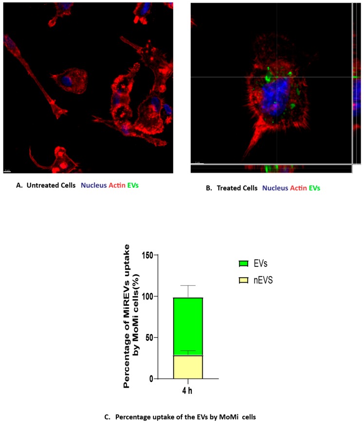
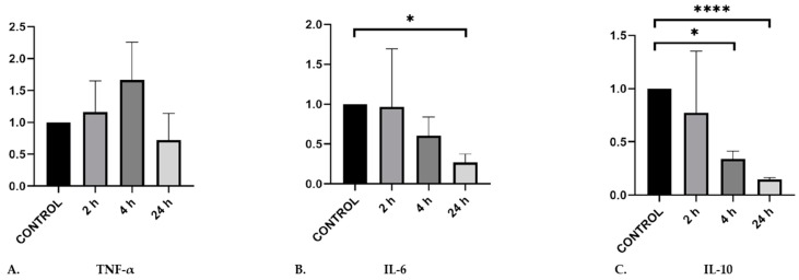
Microglia are the chief immune cells of the brain and have been reported to be activated in severe malaria. Their activation may drive towards neuroinflammation in cerebral malaria. Malaria-infected red blood cell derived-extracellular vesicles (MiREVs) are produced during the blood stage of malaria infection. They mediate intercellular communication and immune regulation, among other functions. During cerebral malaria, the breakdown of the blood-brain barrier can promote the migration of substances such as MiREVs from the periphery into the brain, targeting cells such as microglia. Microglia and extracellular vesicle interactions in different pathological conditions have been reported to induce neuroinflammation. Unlike in astrocytes, microglia-extracellular vesicle interaction has not yet been described in malaria infection. Therefore, in this study, we aimed to investigate the uptake of MiREVs by human microglia cells and their cytokine response. Human blood monocyte-derived microglia (MoMi) were generated from buffy coats of anonymous healthy donors using Ficoll-Paque density gradient centrifugation. The MiREVs were isolated from the Plasmodium falciparum cultures. They were purified by ultracentrifugation and labeled with PKH67 green fluorescent dye. The internalization of MiREVs by MoMi was observed after 4 h of co-incubation on coverslips placed in a 24-well plate at 37 °C using confocal microscopy. Cytokine-gene expression was investigated using rt-qPCR, following the stimulation of the MoMi cells with supernatants from the parasite cultures at 2, 4, and 24 h, respectively. MiREVs were internalized by the microglia and accumulated in the perinuclear region. MiREVs-treated cells increased gene expression of the inflammatory cytokine TNFα and reduced gene expression of the immune suppressive IL-10. Overall, the results indicate that MiREVs may act on microglia, which would contribute to enhanced inflammation in cerebral malaria.
Keywords: extracellular vesicles; malaria-infected red blood cells; microglia.
Conflict of interest statement
There are no potential conflicts of interest with respect to the research, authorship, and/or publication of this article.
Figures
References
-
- World Health Organization . Global Malaria Programme: World Malaria Report 2017. World Health Organization; Geneva, Switzerland: 2017.
LinkOut - more resources
Full Text Sources
