Review
. 2019 Dec 24;25(1):82.
doi: 10.3390/molecules25010082.

Advances in Structure Modeling Methods for Cryo-Electron Microscopy Maps

Affiliations
Review

Advances in Structure Modeling Methods for Cryo-Electron Microscopy Maps

Eman Alnabati et al. Molecules. .

Abstract

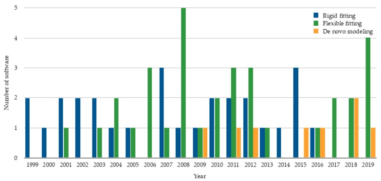
Cryo-electron microscopy (cryo-EM) has now become a widely used technique for structure determination of macromolecular complexes. For modeling molecular structures from density maps of different resolutions, many algorithms have been developed. These algorithms can be categorized into rigid fitting, flexible fitting, and de novo modeling methods. It is also observed that machine learning (ML) techniques have been increasingly applied following the rapid progress of the ML field. Here, we review these different categories of macromolecule structure modeling methods and discuss their advances over time.

Keywords: cryo-EM; cryo-electron microscopy; de novo modeling; density map; machine learning methods; protein modeling; structure fitting algorithms.


Conflict of interest statement

The authors declare no conflict of interest.

Figures

Figure 1
Figure 1
The number of rigid fitting, flexible fitting, and de novo modeling software published per year. The statistics are based on publication. The plot shows 28 rigid fitting methods [9,10,11,12,13,14,15,16,17,18,19,20,21,22,23,24,25,26,27,28,29,30,31,32,33,34,35,36], 33 flexible fitting methods [37,38,39,40,41,42,43,44,45,46,47,48,49,50,51,52,53,54,55,56,57,58,59,60,61,62,63,64,65,66,67,68,69], and 8 de novo modeling methods [70,71,72,73,74,75,76,77].
Figure 2
Figure 2
Schematic flow diagram of the MAINMAST algorithm. The cryo-EM density map shown on the right is of structural protein 5 of cytoplasmic polyhedrosis virus solved at a 2.9 Å resolution (EMD-6374). This figure was adapted from the MAINMAST paper [76].
Figure 3
Figure 3
Emap2sec applied on two experimental maps. Density maps and their fitted protein structures are shown on the left and the secondary structure detection by Emap2sec is shown on the right. Spheres in magenta, yellow, and green show detected α-helices, β-strands, and other structures, respectively. (a) Archaeal 20S proteasome (EMD-1733 of resolution 6.8 Å; PDB 3C91). (b) Eschirichia coli replicative DNA polymerase complex (EMD-3201 of resolution 8.34 Å; PDB 5FKU). This figure was adapted from the EMap2sec paper [101].

References

    1. Cheng Y. Single-Particle Cryo-EM at Crystallographic Resolution. Cell. 2015;161:450–457. doi: 10.1016/j.cell.2015.03.049. - DOI - PMC - PubMed
    1. Nogales E. The development of cryo-EM into a mainstream structural biology technique. Nat. Methods. 2016;13:24. doi: 10.1038/nmeth.3694. - DOI - PMC - PubMed
    1. Glaeser R.M. How good can single-particle cryo-EM become? What remains before it approaches its physical limits? Annu. Rev. Biophys. 2019;48:45–61. - PubMed
    1. Kuhlbrandt W. Biochemistry. The resolution revolution. Science. 2014;343:1443–1444. doi: 10.1126/science.1251652. - DOI - PubMed
    1. Henderson R. Overview and future of single particle electron cryomicroscopy. Arch. Biochem. Biophys. 2015;581:19–24. doi: 10.1016/j.abb.2015.02.036. - DOI - PubMed

LinkOut - more resources