

Retinal hyperreflective foci in Fabry disease
- PMID: 31878969
- PMCID: PMC6933914
- DOI: 10.1186/s13023-019-1267-2
Retinal hyperreflective foci in Fabry disease
Abstract
Background: Fabry disease (FD) is an X-linked inherited storage disorder caused by deficiency of lysosomal alpha-Galactosidase A. Here we describe new retinal findings in patients with FD assessed by Spectral domain optical coherence tomography (SD-OCT) and their possible clinical relevance.
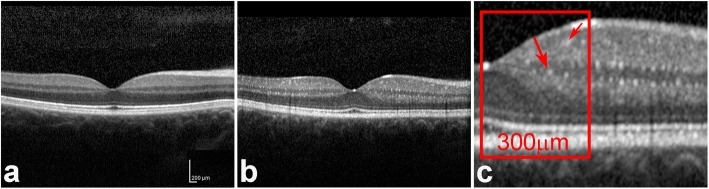
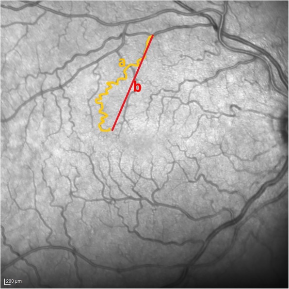
Methods: 54 eyes of 27 FD patients and 54 eyes of 27 control subjects were included. The ophthalmic examination included visual acuity testing, tonometry, slit lamp and fundus examination. SD-OCT imaging of the macula was performed in all subjects. Central retinal thickness and retinal nerve fiber layer analysis were quantified. Vessel tortuosity was obtained by a subjective scoring and mathematically calculated. Inner retinal hyperreflective foci (HRF) were quantified, clinically graded and correlated with a biomarker of Fabry disease (lyso-Gb3).
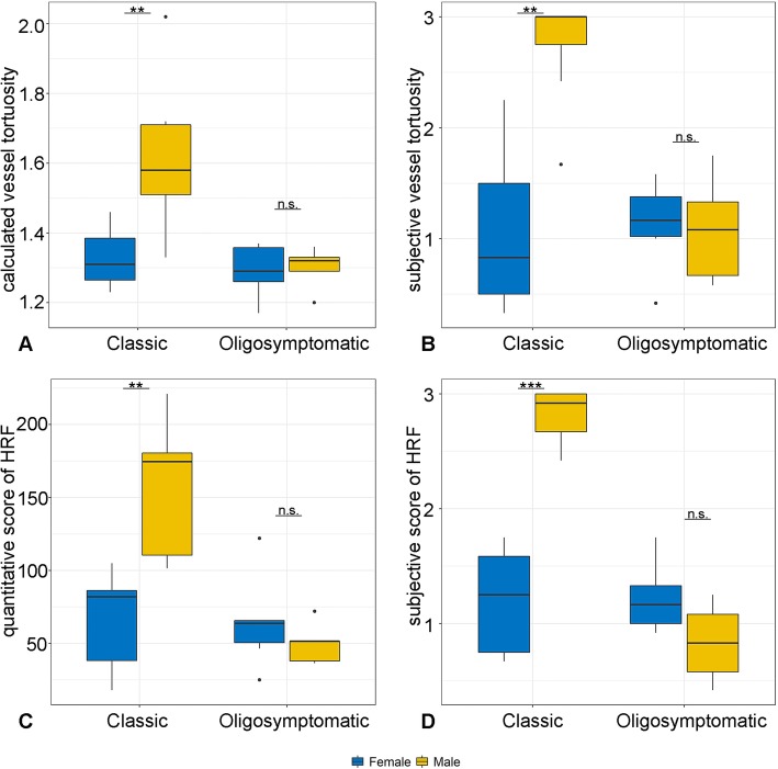
Results: In comparison to an age-matched control group, a significant amount of HRF was identified in macular SD-OCT images in FD patients. These HRF were localized within the inner retinal layers. Furthermore, lyso-Gb3 levels correlated significantly with the quantitative evaluation of HRF (p < 0,001). In addition, the vessel tortuosity was remarkably increased in FD patients compared to control persons and correlated significantly with lyso-G3 levels (p = 0.005). A further subanalysis revealed significantly higher HRF and vessel tortuosity scores in male patients with the classic FD phenotype.
Conclusions: The observational, cross sectional, comparative study describes novel intraretinal findings in patients with FD. We were able to identify suspicious HRF within the inner retinal layers. These findings were not accompanied by functional limitations, as visual acuity remained unchanged. However, HRF correlated well with lyso-Gb3, a degradation product of the accumulating protein Gb3 and might potentially indicate Gb3 accumulation within the highly metabolic and densely vascularized macula.
Keywords: Fabry disease; Hyperreflective foci; Retina; SD-OCT; Vessel tortuosity.
Conflict of interest statement
The authors declare that they have no competing interests
Figures
References
-
- Desnick RJ, Dean KJ, Grabowski GA, Bishop DF, Sweeley CC. Enzyme therapy XVII: metabolic and immunologic evaluation of alpha- galactosidase A replacement in Fabry disease. Birth Defects Orig Artic Ser. 1980;16(1):393–413. - PubMed
Publication types
MeSH terms
Substances
LinkOut - more resources
Full Text Sources
Medical
