Review
. 2020 Feb 14;295(7):1792-1814.
doi: 10.1074/jbc.REV119.009457. Epub 2019 Dec 25.

Membrane receptor activation mechanisms and transmembrane peptide tools to elucidate them

Affiliations
Review

Membrane receptor activation mechanisms and transmembrane peptide tools to elucidate them

Justin M Westerfield et al. J Biol Chem. .

Abstract

Single-pass membrane receptors contain extracellular domains that respond to external stimuli and transmit information to intracellular domains through a single transmembrane (TM) α-helix. Because membrane receptors have various roles in homeostasis, signaling malfunctions of these receptors can cause disease. Despite their importance, there is still much to be understood mechanistically about how single-pass receptors are activated. In general, single-pass receptors respond to extracellular stimuli via alterations in their oligomeric state. The details of this process are still the focus of intense study, and several lines of evidence indicate that the TM domain (TMD) of the receptor plays a central role. We discuss three major mechanistic hypotheses for receptor activation: ligand-induced dimerization, ligand-induced rotation, and receptor clustering. Recent observations suggest that receptors can use a combination of these activation mechanisms and that technical limitations can bias interpretation. Short peptides derived from receptor TMDs, which can be identified by screening or rationally developed on the basis of the structure or sequence of their targets, have provided critical insights into receptor function. Here, we explore recent evidence that, depending on the target receptor, TMD peptides cannot only inhibit but also activate target receptors and can accommodate novel, bifunctional designs. Furthermore, we call for more sharing of negative results to inform the TMD peptide field, which is rapidly transforming into a suite of unique tools with the potential for future therapeutics.

Keywords: T-cell receptor (TCR); cell signaling; conformational change; epidermal growth factor receptor (EGFR); integrin; oligomerization; protein–protein interaction; receptor tyrosine kinase; transmembrane domain; transmembrane receptor; α-helical.


Conflict of interest statement

The authors declare that they have no conflicts of interest with the contents of this article

Figures
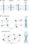
Figure 1.
Figure 1.
Common structural motifs of TMD dimers. A, isolated transmembrane α-helices are constrained to four principal motions: piston movement, translation, tilt, and rotation. B, common characteristics of interhelical interaction motifs. The left-handed TMD dimer is typically found at an approximate −20° crossing angle. Similar to soluble coiled coils, the interaction interface can be described in a heptad repeat nomenclature, abcdefg. Interhelical hydrogen bonding (cyan) is commonly found between a–a′ and d–e′. The average interhelical distance is 11 Å (58). The right-handed TMD dimer typically forms at an approximate +40° crossing angle. Right-handed dimers are best described by a tetrad repeat nomenclature, abcd. At the a and d positions, small residues such as glycine are enriched, which create an angled groove where helices may pack tightly together. Interhelical hydrogen bonding is commonly found between residues at positions a and d′. The average interhelical distance is ∼9 Å (58). Representative structures shown are as follows: left-handed is EphA2 (PDB code 2K9Y), and right-handed is glycophorin A (PDB code 5EH4).
Figure 2.
Figure 2.
Role of TMD in receptor activation mechanisms. The LID hypothesis posits that ligand binding to the extracellular domains of the receptor brings receptor monomers together into a dimer that is signaling-competent. The LIR hypothesis assumes that an inactive dimer exists and that ligand binding induces a rotation of the receptor to bring the intracellular kinase domains into the active configuration for signaling. Clustering occurs when receptors are stabilized as large higher-order oligomeric signaling complexes. These mechanisms are not necessarily mutually exclusive, and some receptors may use a combination of them. Blue and orange represent different TMD interfaces. Domains are not to scale, and the ligand is not shown for clarity.
Figure 3.
Figure 3.
Receptor activation can rely on different processes. LID requires that receptors come into close contact upon ligand binding. The left panel shows that in the presence of ligand, a free energy minimum appears when receptors are in contact (10 Å is the approximate distance from helix centers (58)). In the absence of ligand, receptors will be separated by some context-dependent average distance between monomers. LIR requires reorientation of TMDs. The central panel shows that receptor TMDs may have more than one permitted crossing angle, whereas only one is favored in the presence of ligand. This example shows a ligand-induced shift toward the right-handed (R) conformation. Clustering requires that ligand binding induces formation of larger oligomers. The right panel shows that in the absence of ligand, high-order oligomers are energetically unfavorable, whereas the presence of ligand stabilizes clusters. These processes are not mutually exclusive and may all occur in the same receptor.
Figure 4.
Figure 4.
TMD peptide functional consequences. TMD peptides can have different functional effects based on the receptor activation mechanism. TMD peptides (green) may competitively inhibit receptor dimerization, leading to reduced signaling or, alternatively, increased signaling in the case of integrins. Receptor complexes may also be stabilized by TMD peptides such as the traptamers and TYPE7, leading to activation, although how this occurs structurally is still unclear. TMD peptides interact preferentially with a specific interface of the TMD shown in blue.

References

    1. Sigalov A. B. (2010) New therapeutic strategies targeting transmembrane signal transduction in the immune system. Cell Adh. Migr. 4, 255–267 10.4161/cam.4.2.10746 - DOI - PMC - PubMed
    1. Stone T. A., and Deber C. M. (2017) Therapeutic design of peptide modulators of protein–protein interactions in membranes. Biochim. Biophys. Acta 1859, 577–585 10.1016/j.bbamem.2016.08.013 - DOI - PubMed
    1. Yin H., and Flynn A. D. (2016) Drugging membrane protein interactions. Annu. Rev. Biomed. Eng. 18, 51–76 10.1146/annurev-bioeng-092115-025322 - DOI - PMC - PubMed
    1. Zeng X., Wu P., Yao C., Liang J., Zhang S., and Yin H. (2017) Small molecule and peptide recognition of protein transmembrane domains. Biochemistry 56, 2076–2085 10.1021/acs.biochem.6b00909 - DOI - PubMed
    1. Zhao T. X., Martinko A. J., Le V. H., Zhao J., and Yin H. (2010) Development of agents that modulate protein–protein interactions in membranes. Curr. Pharm. Des. 16, 1055–1062 10.2174/138161210790963878 - DOI - PubMed

Publication types

MeSH terms