. 2020 Jan 14;117(2):1139-1147.
doi: 10.1073/pnas.1912109117. Epub 2019 Dec 26.

The circadian clock protein REVERBα inhibits pulmonary fibrosis development

Affiliations

The circadian clock protein REVERBα inhibits pulmonary fibrosis development

Peter S Cunningham et al. Proc Natl Acad Sci U S A. .

Abstract

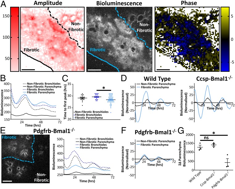
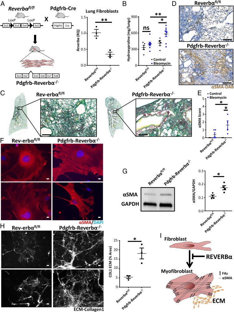
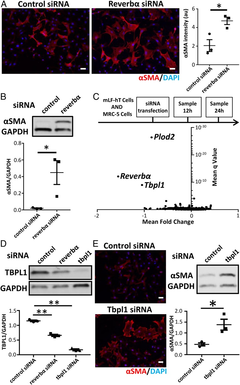
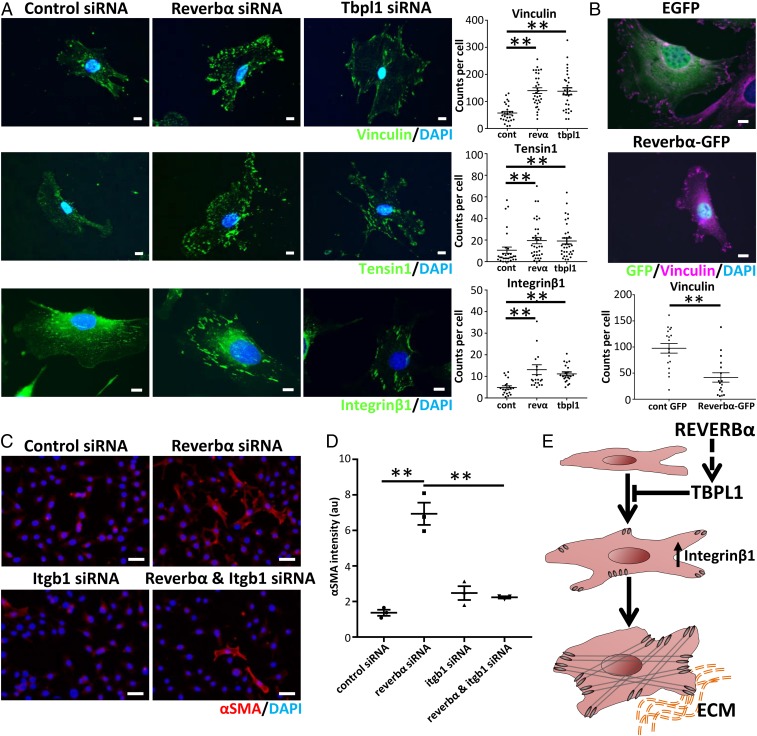
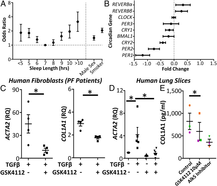
Pulmonary inflammatory responses lie under circadian control; however, the importance of circadian mechanisms in the underlying fibrotic phenotype is not understood. Here, we identify a striking change to these mechanisms resulting in a gain of amplitude and lack of synchrony within pulmonary fibrotic tissue. These changes result from an infiltration of mesenchymal cells, an important cell type in the pathogenesis of pulmonary fibrosis. Mutation of the core clock protein REVERBα in these cells exacerbated the development of bleomycin-induced fibrosis, whereas mutation of REVERBα in club or myeloid cells had no effect on the bleomycin phenotype. Knockdown of REVERBα revealed regulation of the little-understood transcription factor TBPL1. Both REVERBα and TBPL1 altered integrinβ1 focal-adhesion formation, resulting in increased myofibroblast activation. The translational importance of our findings was established through analysis of 2 human cohorts. In the UK Biobank, circadian strain markers (sleep length, chronotype, and shift work) are associated with pulmonary fibrosis, making them risk factors. In a separate cohort, REVERBα expression was increased in human idiopathic pulmonary fibrosis (IPF) lung tissue. Pharmacological targeting of REVERBα inhibited myofibroblast activation in IPF fibroblasts and collagen secretion in organotypic cultures from IPF patients, thus suggesting that targeting of REVERBα could be a viable therapeutic approach.

Keywords: Reverb alpha; circadian; integrin; pulmonary fibrosis; sleep.


Conflict of interest statement

The authors declare no competing interest.

Figures

Fig. 1.
Fig. 1.
Asynchronous circadian oscillations occur in pulmonary fibrosis. (A) Bioluminescent image along with heat maps of amplitude and phase taken from the same PCLS obtained from a mPER2::luc mouse 14 d after in vivo bleomycin treatment (3 U/kg). Data are representative of 3 separate experiments. (Scale bars, 500 µm.) (B) Bioluminescent intensity plotted against time for both parenchyma and bronchioles in fibrotic and nonfibrotic regions (data are representative of 3 separate experiments). (C) Time to first peak for bronchioles and parenchyma in fibrotic and nonfibrotic areas. *P < 0.05 (ANOVA with post hoc Dunnett test using 18, 19, and 48 representative sections for healthy airways, fibrotic airways, and fibrotic parenchyma, respectively, in the lung slice). Data are representative of 3 separate experiments (mean ± SEM). (D) Bioluminescent intensity plotted against time (24-h moving average baseline subtracted) for the representative slices shown in A and Ccsp-Bmal1−/− mice shown in SI Appendix, Fig. S2A. (E) Representative bioluminescent image along with bioluminescent intensity plotted against time for a PCLS 14 d after in vivo bleomycin treatment in the Pdgfrb-Bmal1−/− mPER2::luc mouse (3 U/kg). Data are representative of 3 separate experiments. (Scale bar, 500 µm.) (F) Bioluminescent intensity plotted against time (24-h moving average baseline subtracted) for the Pdgfrb-Bmal1−/− representative slice shown in E. (G) Difference in bioluminescence between fibrotic and nonfibrotic parenchyma over 3 d in PCLS from WT, Ccsp-Bmal1−/−, and Pdgfrb-Bmal1−/− mice after in vivo bleomycin treatment (n = 3 animals). ns, not significant. *P < 0.05 (1-way ANOVA Dunnett post hoc test; mean ± SEM).
Fig. 2.
Fig. 2.
REVERBα alters susceptibility to pulmonary fibrosis through its effect on myofibroblast differentiation. (A) Schematic showing generation of Pdgfrb-Reverbα−/− mice combined with qPCR analysis of Reverbα expression in lung fibroblasts (n = 3 animals). **P < 0.01 (Student t test; mean ± SEM). (B) Hydroxyproline measurement in lungs from Pdgfrb-Reverbα−/− mice and littermate controls 28 d following challenge with intratracheal bleomycin (2 U/kg) or saline (n = 4 to 5 saline and 8 bleomycin per genotype). *P < 0.05; **P < 0.01 (2-way ANOVA Holm–Sidak post hoc test; mean ± SEM). (C) In a separate experiment, histology (Picrosirius red) of lungs was examined 28 d following challenge with intratracheal bleomycin (representative image from 4 animals treated with bleomycin per genotype). (Scale bars, 200 µm.) (D) Immunohistochemical staining of myofibroblasts (anti-αSMA, 3,3′-diaminobenzidine [DAB]) from Pdgfrb-Reverbα−/− mice and littermate controls 28 d following intratracheal bleomycin challenge (representative image from 4 animals treated with bleomycin per genotype). (Scale bars, 200 µm.) (E) Histological scoring (grade 0 to 4) for the presence of αSMA staining 28 d following intratracheal bleomycin challenge (n = 3 saline and 4 to 5 bleomycin per genotype). *P < 0.05 (2-way ANOVA Holm–Sidak post hoc test; mean ± SEM). (F and G) Representative immunofluorescence images of primary lung fibroblast cultures from Pdgfrb-Reverbα−/− mice and littermate controls showing intracellular αSMA (red) (n = 3 animals per genotype) (F) combined with a representative immunoblot and quantification of intracellular αSMA from primary lung fibroblast cultures (n = 4 animals per genotype) (G). *P < 0.05 (Student t test; mean ± SEM). DAPI, 4′,6-diamidino-2-phenylindole. (Scale bars in F, 10 µm.) (H) Representative collagen-1 ECM (extracellular matrix) images and quantification following culture of Pdgfrb-Reverbα−/− and Reverbαfl/fl primary lung fibroblasts (n = 3 animals per genotype). *P < 0.05 (Student t test; mean ± SEM). (Scale bars, 50 µm.) (I) Schematic illustrating the action of REVERBα in inhibiting fibroblast/myofibroblast differentiation. FAs, focal adhesions.
Fig. 3.
Fig. 3.
REVERBα alters myofibroblast differentiation via TBPL1. (A) Immunofluorescent staining and quantification for the myofibroblast marker αSMA after control (nontargeting) or Reverbα siRNA knockdown in mLF-hT cells (n = 3 separate transfections). *P < 0.05 (Student t test; mean ± SEM). (Scale bars, 50 µm.) (B) Immunoblot and densitometry for αSMA in MRC-5 cells after control (nontargeting) or REVERBα siRNA knockdown (representative immunoblot shown; n = 3 separate transfections). *P < 0.05 (Student t test; mean ± SEM). (C) Schematic of RNA-seq sample preparation. Control (nontargeting) or Reverbα siRNA knockdown was performed in 2 fibroblast cell lines (mLF-hT cells and MRC-5). Samples were collected for RNA-seq analysis 12 and 24 h after siRNA transfection from 3 separate transfections for each cell line per time point. Pooled analysis of all 4 different RNA-seq experimental conditions is shown by a volcano plot (mean fold change plotted against mean q-value). (D) Immunoblot of TBPL1 following control (nontargeting) or Reverbα or Tbpl1 siRNA knockdown in mLF-hT cells (a representative immunoblot is shown; n = 3 separate transfections). **P < 0.01 (Student t test; mean ± SEM). (E) Representative immunofluorescence and immunoblotting for αSMA after control (nontargeting) or Tbpl1 siRNA knockdown in mLF-hT cells (a representative immunoblot is shown; n = 3 separate transfections). *P < 0.05 (Student t test; mean ± SEM). (Scale bars, 50 µm.) DAPI, 4′,6-diamidino-2-phenylindole.
Fig. 4.
Fig. 4.
REVERBα and TBPL1 affect myofibroblast differentiation through changes in integrinβ1 expression. (A) Representative immunofluorescent images and quantification per cell of vinculin, tensin1, and integrinβ1 following siRNA knockdown of Reverbα or Tbpl1 compared to control (nontargeting) siRNA in mLF-hT cells (n = 3 separate transfections). **P < 0.01 (1-way ANOVA post hoc Dunnett test; mean ± SEM). Dots represent individual cells from 3 transfections. cont, control siRNA; revα, Reverbα siRNA. (Scale bars, 10 µm.) (B) Representative immunofluorescence image after mLF-hT cells have been transfected with REVERBα-GFP plasmid or an empty-GFP plasmid. Cells were stained for GFP, vinculin, and nuclei (4′,6-diamidino-2-phenylindole [DAPI]) (n = 3 separate transfections). **P < 0.01 (Student t test; mean ± SEM). Dots represent individual cells from 3 transfections with the focal-adhesion number being quantified per cell. (Scale bars, 10 µm.) (C) Representative immunofluorescence images. (Scale bars, 50 µm.) (D) Quantification of the myofibroblast marker αSMA following dual siRNA knockdown (control or Reverbα in the presence or absence of Itgb1) in mLF-hT cells (n = 3 separate transfections). **P ≤ 0.01 (1-way ANOVA post hoc Dunnett test; mean ± SEM). (E) Schematic demonstrating how both REVERBα and TBPL1 regulate Integrinβ1, which in turn affects myofibroblast differentiation. ECM, extracellular matrix.
Fig. 5.
Fig. 5.
Circadian factors are associated with IPF, where a REVERB ligand represses collagen secretion. (A) Odds ratio (OR) for the association between pulmonary fibrosis and sleep duration (OR ± 95% confidence interval (CI); logistic regression; n = 500,074 subjects from the UK Biobank). (B) Changes in clock-gene expression in IPF compared to control subjects from a previously published genome array (GSE47460) (fold change ± 95% confidence interval; n = 90 controls and 98 patients with IPF). (C) qPCR for αSMA (ACTA2) and Collagen1 (COL1A1) following TGFβ stimulation (2 ng/mL) in primary human lung fibroblasts obtained from patients with pulmonary fibrosis in the presence or absence of GSK4112 (10 μM) (n = 4 fibrotic patients). *P < 0.05 (Student t test; mean ± SEM). (D) qPCR for αSMA (ACTA2) expression following treatment with TGFβ (2 ng/mL) and GSK4112 (10 μM) in human PCLS (n = 5 patients). *P < 0.05 (Student t test; mean ± SEM). (E) Enzyme-linked immunosorbent assay analysis of secreted collagen-1 in TGFβ-stimulated PCLS obtained from 3 patients with IPF treated with the REVERB ligand GSK4112 (10 μM) and an Alk5 inhibitor (1 μM) as positive control (n = 3). *P < 0.05 (paired Student t test; mean ± SEM).

Comment in

References

    1. Pariollaud M., et al. , Circadian clock component REV-ERBα controls homeostatic regulation of pulmonary inflammation. J. Clin. Invest. 128, 2281–2296 (2018). - PMC - PubMed
    1. Gibbs J., et al. , An epithelial circadian clock controls pulmonary inflammation and glucocorticoid action. Nat. Med. 20, 919–926 (2014). - PMC - PubMed
    1. Cunningham P. S., et al. , Incidence of primary graft dysfunction after lung transplantation is altered by timing of allograft implantation. Thorax 74, 413–416 (2019). - PMC - PubMed
    1. Pekovic-Vaughan V., et al. , The circadian clock regulates rhythmic activation of the NRF2/glutathione-mediated antioxidant defense pathway to modulate pulmonary fibrosis. Genes Dev. 28, 548–560 (2014). - PMC - PubMed
    1. Gibbs J. E., et al. , The nuclear receptor REV-ERBα mediates circadian regulation of innate immunity through selective regulation of inflammatory cytokines. Proc. Natl. Acad. Sci. U.S.A. 109, 582–587 (2012). - PMC - PubMed

Publication types

MeSH terms

LinkOut - more resources