

Clinicopathological and ultrastructural characterization of periapical actinomycosis
- PMID: 31880281
- PMCID: PMC6982982
- DOI: 10.4317/medoral.23247
Clinicopathological and ultrastructural characterization of periapical actinomycosis
Abstract
Background: The aim of the present study was to analyze the clinicopathological and the ultrastructural features of periapical actinomycosis (PA) cases.
Material and methods: Data from the files of an oral pathology laboratory were retrieved and the findings of histopathological analysis were evaluated. Hematoxylin-eosin (HE), a modified Brown & Brenn, and Grocott stains as well as ultrastructural analysis using scanning electron microscopy (SEM) and energy dispersive X-ray spectroscopy (EDX) were utilized.
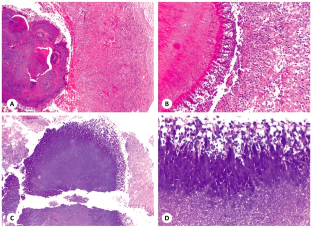
Results: Six cases were obtained, 4 females and 2 males, with a mean age of 34 year-old. Two cases were symptomatic, lower teeth and the anterior region were more commonly affected, and all cases were characterized by periapical radiolucencies. All cases presented sulfur granules with a ray-fungus or club-shaped pattern of the Splendore-Hoeppli phenomenon in HE-stained sections, with filamentous gram-positive bacteria aggregates highlighted by the modified Brown & Brenn stain. SEM analysis revealed abundant packed rod-like and filamentous bacteria associated with an extracellular amorphous material. EDX analysis showed predominant picks of calcium and sulfur in actinomycotic colonies.
Conclusions: Our findings suggest that PA manifests either clinically and radiologically as a non-specific and heterogeneous condition and that the actinomycotic colonies consist in a calcium- and sulfur-rich matrix. Furthermore, the results highlight the importance of submitting periapical specimens after surgical removal to histopathological analysis.
Conflict of interest statement
Conflicts of interest The authors deny any conflicts of interest related to this study. We affirm that we have no financial affiliation (e.g., employment, direct payment, stock holdings, retainers, consultantships, patent licensing arrangements or honoraria), or involvement with any commercial organization with direct financial interest in the subject or materials discussed in this manuscript, nor have any such arrangements existed in the past three years. The authors deny any conflicts of interest related to this study.
Figures


References
-
- Wong VK, Turmezei TD, Weston VC. Actinomycosis. BMJ. 2011;343:d6099. - PubMed
-
- Brown JR. Human actinomycosis: a study of 181 subjects. Hum Pathol. 1973;4:319–30. - PubMed
-
- Israel J. Neve Beobactungen anf dem Bebiete der Mykosen des Menshen. Virchows Arch Pathol Anat Physiol Klin Med. 1878;74:15–53.
-
- Wolfe M, Israel J. Ueber Reincultur des Actinomyces and seine Uebertragbarkeit auf Thiere. Virchows Arch Pathol Anat Physiol Klin Med. 1891;126:11–59.
-
- Lerner PI. The lumpy jaw. Cervicofacial actinomycosis. Infect Dis Clin North Am. 1988;2:203–20. - PubMed
MeSH terms
LinkOut - more resources
Full Text Sources
