. 2020 Feb;24(3):2319-2329.
doi: 10.1111/jcmm.14914. Epub 2019 Dec 27.

Up-regulation of paired-related homeobox 2 promotes cardiac fibrosis in mice following myocardial infarction by targeting of Wnt5a

Affiliations

Up-regulation of paired-related homeobox 2 promotes cardiac fibrosis in mice following myocardial infarction by targeting of Wnt5a

Wen-Wu Bai et al. J Cell Mol Med. 2020 Feb.

Abstract

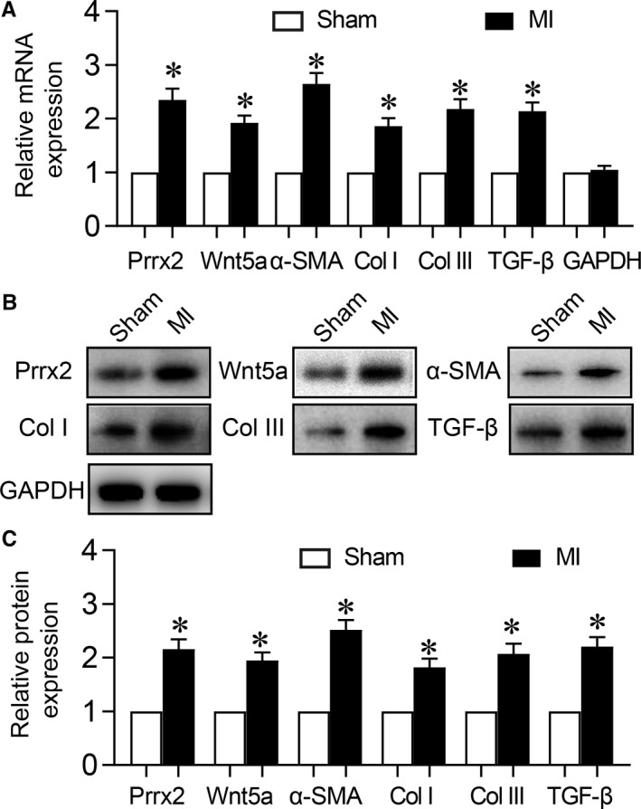
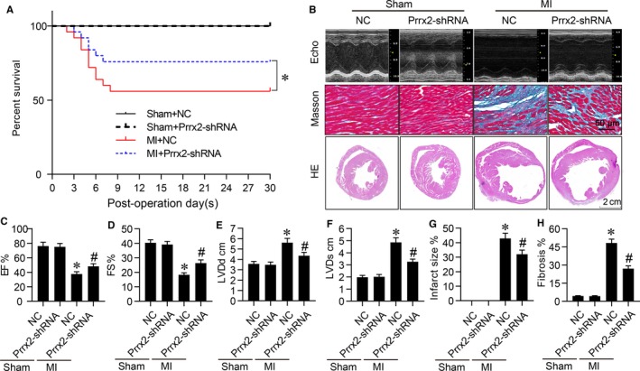
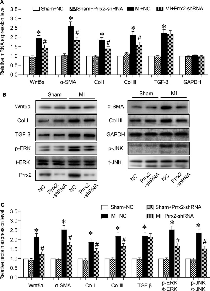
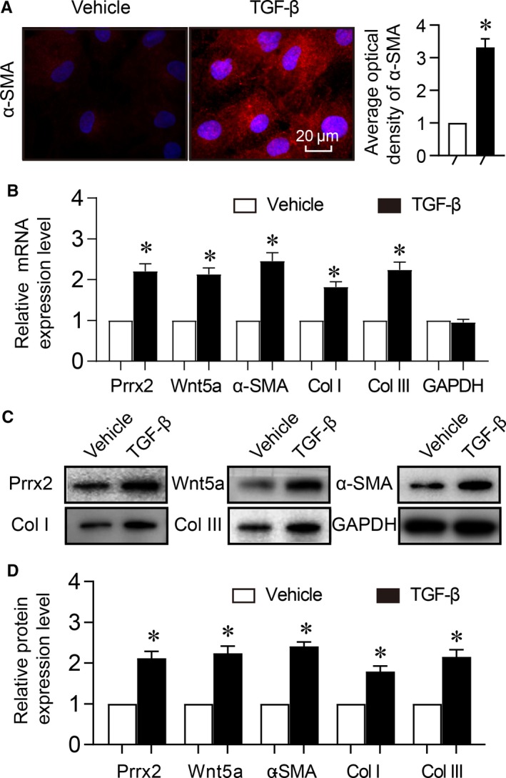
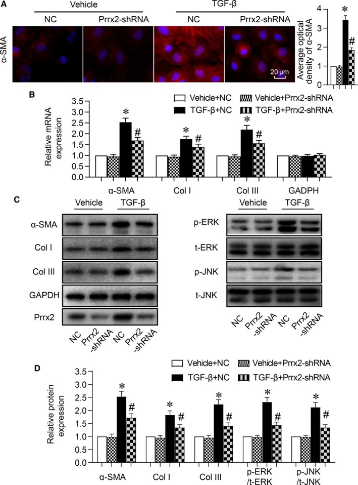
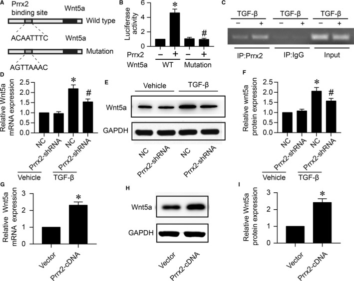
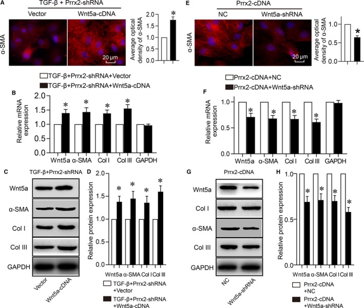
Cardiac fibrosis is a key factor to determine the prognosis in patient with myocardial infarction (MI). The aim of this study is to investigate whether the transcriptional factor paired-related homeobox 2 (Prrx2) regulates Wnt5a gene expression and the role in myocardial fibrosis following MI. The MI surgery was performed by ligation of left anterior descending coronary artery. Cardiac remodelling was assessed by measuring interstitial fibrosis performed with Masson staining. Cell differentiation was examined by analysis the expression of alpha-smooth muscle actin (α-SMA). Both Prrx2 and Wnt5a gene expressions were up-regulated in mice following MI, accompanied with increased mRNA and protein levels of α-SMA, collagen I and collagen III, compared to mice with sham surgery. Adenovirus-mediated gene knock down of Prrx2 increased survival rate, alleviated cardiac fibrosis, decreased infarction sizes and improved cardiac functions in mice with MI. Importantly, inhibition of Prrx2 suppressed ischaemia-induced Wnt5a gene expression and Wnt5a signalling. In cultured cardiac fibroblasts, TGF-β increased gene expressions of Prrx2 and Wnt5a, and induced cell differentiations, which were abolished by gene silence of either Prrx2 or Wnt5a. Further, overexpression of Prrx2 or Wnt5a mirrored the effects of TGF-β on cell differentiations of cardiac fibroblasts. Gene silence of Wnt5a also ablated cell differentiations induced by Prrx2 overexpression in cardiac fibroblasts. Mechanically, Prrx2 was able to bind with Wnt5a gene promoter to up-regulate Wnt5a gene expression. In conclusions, targeting Prrx2-Wnt5a signalling should be considered to improve cardiac remodelling in patients with ischaemic heart diseases.

Keywords: Wnt5a; cardiac fibrosis; cell differentiation; myocardial infarction; paired-related homeobox 2.


Conflict of interest statement

None.

Figures

Figure 1
Figure 1
Ischaemia up‐regulates Prrx2 and Wnt5a gene expressions in mice following MI. Male Apoe−/− mice were subjected to perform MI surgery and hearts were isolated from mice at the 30th post‐operative days to detect (A) gene expressions of Prrx2, Wnt5a, α‐SMA, GAPDH, collagen I (Col I) and collagen III (Col III) by real‐time PCR. B and C, Protein levels of Prrx2, Wnt5a, α‐SMA, GAPDH, collagen I and collagen III in heart homogenates were determined by Western blotting in B and quantitative analysis was performed in C. N is 10‐15 in each group. *P < .05 vs Sham
Figure 2
Figure 2
Gene knockdown of Prrx2 inhibits cardiac fibrosis and improves heart functions in mice after MI. Male Apoe−/− mice were injected with adenovirus expressing negative control (NC) shRNA or Prrx2 shRNA for 3 d prior to MI surgery. At the 30th post‐operative day, mice were subjected to assess cardiac functions by echocardiography before sacrificed and hearts were isolated to measure infraction sizes by HE staining and collagens by Masson staining. A, The survival curve of mice within 30 d after MI surgery. B, Representative images of HE staining, Masson staining and cardiac functions in hearts were shown. C‐F, Cardiac functions were quantified by calculating EF in C, FS in D, measuring LVDd in E and LVDs in F. G, Quantitative analysis of infarct size was performed. H, Quantitative analysis of cardiac fibrosis was conducted. N is 10‐15 in each group. *P < .05 vs NC plus Sham. # P < .05 vs NC plus MI
Figure 3
Figure 3
Adenovirus‐mediated Prrx2 shRNA expression down‐regulates Wnt5a signalling in ischaemic hearts in mice. Male Apoe−/− mice were injected with adenovirus expressing negative control (NC) shRNA or Prrx2 shRNA for 3 d prior to MI surgery. At the 30th post‐operative day, mice were killed under anaesthesia and hearts were isolated to measure (A) gene expressions of Wnt5a, α‐SMA, GAPDH, collagen I (Col I) and collagen III (Col III) by real‐time PCR. B and C, Protein levels of Wnt5a, α‐SMA, TGF‐β, p‐ERK, p‐JNK, collagen I and collagen III in isolated hearts from mice were determined by Western blotting in B and quantitative analysis was performed in C. N is 10‐15 in each group. *P < .05 vs NC plus Sham. # P < .05 vs NC plus MI
Figure 4
Figure 4
TGF‐β induces cell differentiation and up‐regulates Prrx2 and Wnt5a in cardiac fibroblasts. Cultured cardiac fibroblasts were treated with TGF‐β (10 ng/mL) for 24 h. A, Cell differentiation was determined by immunofluorescence analysis of α‐SMA and quantitative analysis was shown. B, Gene expressions of Prrx2, Wnt5a, α‐SMA, GAPDH, collagen I (Col I) and collagen III (Col III) in cells were measured by real‐time PCR. C and D, Total cell lysates of cardiac fibroblasts were subjected to perform Western blotting analysis to detect protein levels of Prrx2, Wnt5a, α‐SMA, Col I and Col III in C and quantitative analysis was performed in D. N is 5 in each group. *P < .05 vs Vehicle
Figure 5
Figure 5
Cell differentiation induced by TGF‐β is abolished by gene silence of Prrx2 in cardiac fibroblasts. Cultured cardiac fibroblasts were infected with adenovirus expressing negative control (NC) shRNA or Prrx2 shRNA for 48 h and then treated with TGF‐β (10 ng/mL) for 24 h. A, Cell differentiation was determined by immunofluorescence analysis of α‐SMA and quantitative analysis was shown. B, Gene expressions of α‐SMA, GAPDH, collagen I (Col I) and collagen III (Col III) in cells were measured by real‐time PCR. C and D, Total cell lysates of cardiac fibroblasts were subjected to perform Western blotting analysis to detect protein levels of α‐SMA, p‐ERK, p‐JNK, Col I, and Col III in C and quantitative analysis was performed in D. N is 5 in each group. *P < .05 vs NC plus Vehicle. # P < .05 vs NC plus TGF‐β
Figure 6
Figure 6
Prrx2 functions as a transcriptional factor of Wnt5a which is up‐regulated by TGF‐β. A, The prediction of binding site for Prrx2 in Wnt5a promoter. B, HEK293A cells were cotransfected with pGL3‐promotor vector constructs expressing WT‐Wnt5a‐promoter containing ACAATTTC or MT‐Wnt5a‐promoter including AGTTAAAC plus Prrx2 cDNA plasmid. Cells were subjected to detect the relative luciferase activity. N is 5 per group. *P < .05 vs WT‐Wnt5a‐promoter. # P < .05 vs WT‐Wnt5a‐promoter plus Prrx2. C, Cultured cardiac fibroblasts were treated with TGF‐β (10 ng/mL) for 24 h. Cells were subjected to detect the binding of Prrx2 to Wnt5a gene promoter by using ChIP method. For ChIP experiment, complex of chromatin/protein was pulled down by Prrx2 primary antibody. The promoter of Wnt5a was amplified by PCR. Positive control is the 10% of the total chromatin in the absence of immunoprecipitation. Negative control is the chromatin immunoprecipitated with IgG and amplified with Wnt5a promoter primers. The PCR production is 200 bp. D‐F, Cultured cardiac fibroblasts were infected with adenovirus expressing negative control (NC) shRNA or Prrx2 shRNA for 48 h and then treated with TGF‐β (10 ng/mL) for 24 h. Total cell lysates were subjected to detect gene expression of Wnt5a by real‐time PCR in D and protein level of Wnt5a by Western blotting in E. Quantitative analysis of Wnt5a protein level was performed in F. N is 5 in each group. *P < .05 vs NC plus Vehicle. # P < .05 vs NC plus TGF‐β. G‐I, Cardiac fibroblasts were infected with adenovirus vector or expressing Prrx2 cDNA for 48 h. Cells were subjected to detect Wnt5a gene expression in G and protein level in H, and quantitative analysis of Wnt5a protein level was shown in I. N is 5 in each group. *P < .05 vs Vector
Figure 7
Figure 7
TGFβ1 via Prrx2‐Wnt5a signalling induces cell differentiation in cardiac fibroblasts. A‐D, Cultured cardiac fibroblasts were co‐infected with adenovirus expressing Prrx2 shRNA plus vector or Wnt5a cDNA for 48 h and then treated with TGF‐β (10 ng/mL) for 24 h. Cell differentiation was determined by immunofluorescence analysis of α‐SMA and quantitative analysis was shown in A. Gene expressions of Wnt5a, α‐SMA, GAPDH, collagen I (Col I) and collagen III (Col III) in cells were measured by real‐time PCR in B. Total cell lysates of cardiac fibroblasts were subjected to perform Western blotting analysis to detect protein levels of Wnt5a, α‐SMA, Col I, and Col III in C and quantitative analysis was performed in D. N is 5 in each group. *P < .05 vs Vector. E‐H, Cardiac fibroblasts were co‐infected with adenovirus expressing Prrx2 cDNA plus negative control (NC) shRNA or Wnt5a shRNA for 48 h. Cells were subjected to detect immunofluorescence analysis of α‐SMA and quantitative analysis was shown in E, gene expressions of Wnt5a, α‐SMA, GAPDH, Col I and Col III in F, protein levels of Wnt5a, α‐SMA, Col I, and Col III in G and H. N is 5 in each group. *P < .05 vs NC plus Prrx2

References

    1. Carney RM, Freedland KE. Depression and coronary heart disease. Nat Rev Cardiol. 2017;14:145‐155. - PubMed
    1. Zhang HM, Liu MY, Lu JX, et al. Intracellular acidosis via activation of Akt‐Girdin signaling promotes post ischemic angiogenesis during hyperglycemia. Int J Cardiol. 2019;277:205‐211. - PubMed
    1. Souders CA, Bowers SL, Baudino TA. Cardiac fibroblast: the renaissance cell. Circ Res. 2009;105:1164‐1176. - PMC - PubMed
    1. Travers JG, Kamal FA, Robbins J, Yutzey KE, Blaxall BC. Cardiac fibrosis: the fibroblast awakens. Circ Res. 2016;118:1021‐1040. - PMC - PubMed
    1. Pan SC, Cui HH, Qiu CG. HOTAIR promotes myocardial fibrosis through regulating URI1 expression via Wnt pathway. Eur Rev Med Pharmacol Sci. 2018;22:6983‐6990. - PubMed

Publication types

MeSH terms