Review
. 2020 Apr;38(4):404-417.
doi: 10.1016/j.tibtech.2019.11.005. Epub 2019 Dec 25.

Natural Medicinal Compounds in Bone Tissue Engineering

Affiliations
Review

Natural Medicinal Compounds in Bone Tissue Engineering

Susmita Bose et al. Trends Biotechnol. 2020 Apr.

Abstract

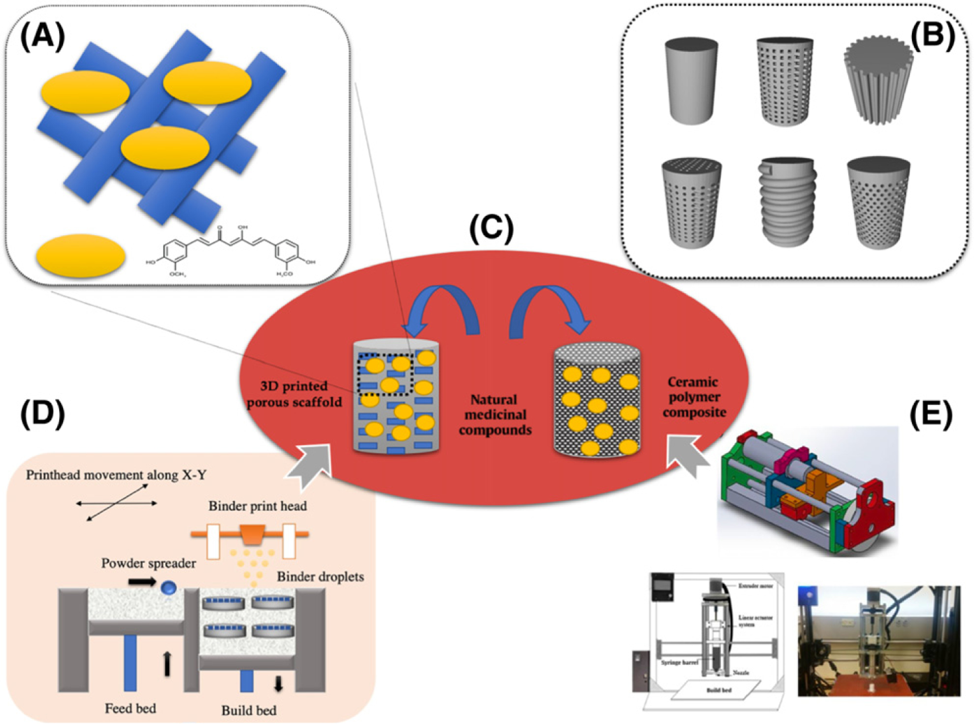
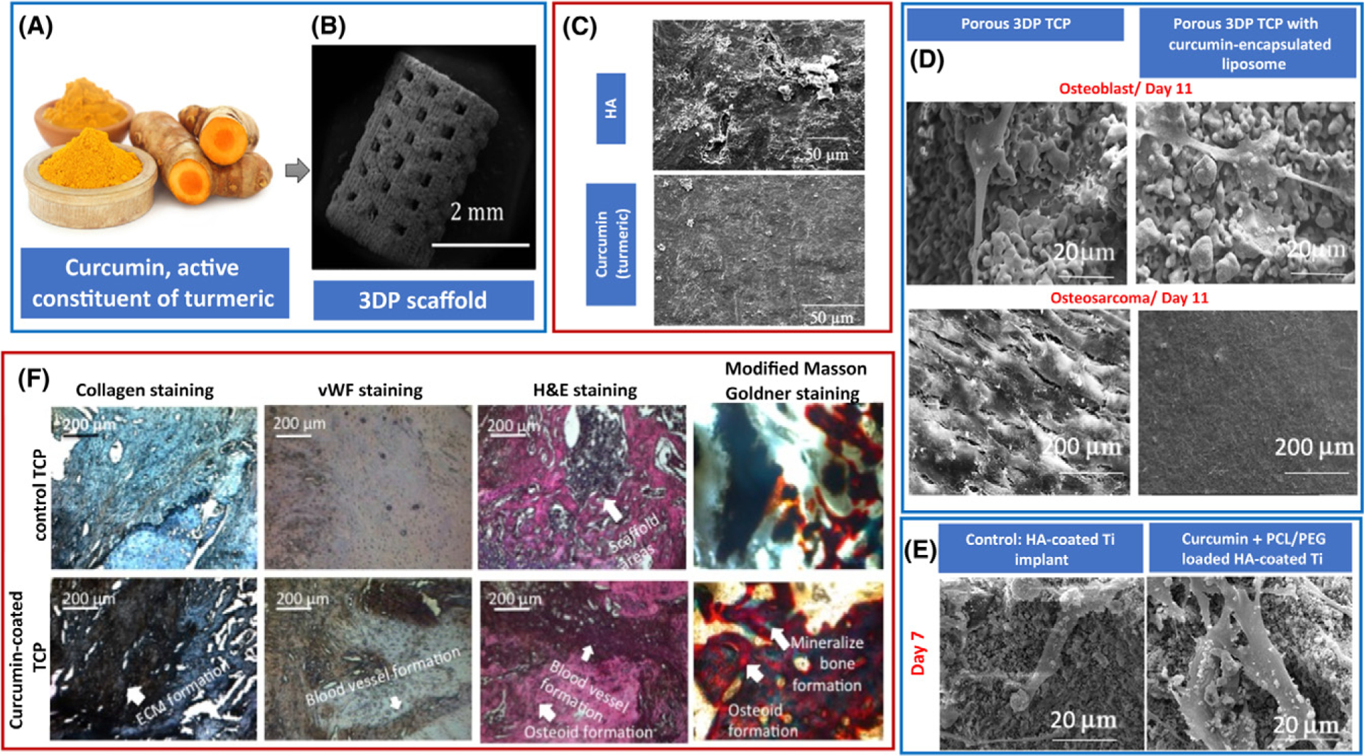
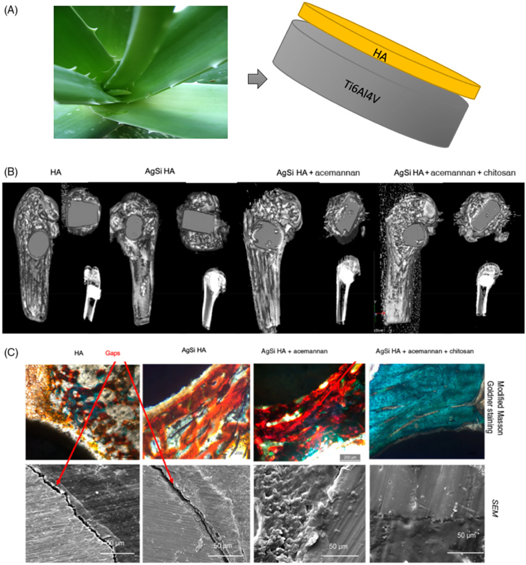
Recent advances in 3D printing have provided unprecedented opportunities in bone tissue engineering applications for producing a variety of complex patient-specific implants for the treatment of critical-sized bone defects. Natural medicinal compounds (NMCs) with osteogenic potential can be incorporated into these 3D-printed parts to improve bone formation and therefore enhance implant performance. Using NMCs to treat bone-related disorders may prove to be a healthy preventive choice as they are considered safe, have lesser or no side effects, and are more suitable for prolonged use than synthetic drugs. In this review paper, the current challenges of bone tissue engineering are addressed briefly, highlighting the immense potential of NMCs integrated within tissue engineering scaffolds for orthopedic and dental applications.

Keywords: 3D printing; bone graft substitutes; bone tissue engineering; drug delivery; natural medicinal compounds.


Figures

Figure 1.
Figure 1.
Natural medicinal compounds can be entrapped within an appropriate delivery vehicle followed by incorporation in a bone scaffold to promote bone regeneration and enhance defect repair.
Figure 2.
Figure 2.. Integration Tools for Natural Medicinal Compounds and Bone Tissue Scaffold.
Schematic illustration showing novel integration approaches of natural medicinal compounds and additively manufactured bone scaffolds.
Figure 3.
Figure 3.. Effects of Curcumin Incorporated 3D Printed Scaffold in Bone Tissue Engineering.
(A) Curcumin, the chief active ingredient from turmeric. (B) Curcumin-coated 3D-printed (3DP) tricalcium phosphate (TCP) scaffolds with designed pores ranging from 340 to 350 μm. (C) Much lesser resorption pits in curcumin-loaded bone implants indicating lower osteoclast activity and therefore reduced bone resorption. (D) Effects of curcumin-encapsulated liposome-coated 3DP scaffolds on osteoblast (healthy bone cell) and osteosarcoma (bone cancer) cells after 11 days of culture; curcumin showed cytotoxicity against osteosarcoma cells by inhibiting the cellular proliferation on the scaffold surface. However, presence of curcumin-encapsulated liposome promoted osteoblast cell attachment and proliferation. (E) Field emission scanning electron microscopy images showing enhanced osteoblast cell attachment and proliferation in presence of curcumin + poly-ε-caprolactone (PCL)/polyethylene glycol (PEG) samples after 7 days of culture. (F) Optical microscopy images of tissue-implant sections after von Willebrand Factor (vWF), hematoxylin and eosin (H&E), collagen, and modified Masson Goldner trichrome staining showing angiogenesis and osteoid-like new bone formation, respectively after 6 weeks of surgery in rat distal femur model. Image reproduced, with permission, from [58,59]. Abbreviations: ECM, Extracellular matrix; HA, hydroxyapatite.
Figure 4.
Figure 4.. Effects of Aloe vera Extract on In Vivo and In Vitro Biological Properties of HA-coated Ti Load-Bearing Implants
. (A) Acemannan, key component of Aloe vera incorporated hydroxyapatite (HA)-coated Ti implant. (B) Computed tomography scan, (B) histology and scanning electron microscopy (SEM) micrographs of HA-coated Ti64 implants with or without dopants and acemannan. The presence of acemannan improved the osteoid formation, which is evident from the enhanced reddish orange color around the vicinity of the implant. SEM micrographs also showed no gaps at the interface of the acemannan implants. The combined presence of chitosan with acemannan further mineralized the new osteoid formation around the implant, shown by the greenish blue color, leading to improved early stage osseointegration. Osseous tissue had been observed to interlock with the implant in the SEM micrographs, leaving no visible gaps. Image adapted, with permission, from [68].

References

    1. Amini AR et al. (2012) Bone tissue engineering: recent advances and challenges. Crit. Rev. Biomed. Eng 40, 363–408 - PMC - PubMed
    1. Younger EM and Chapman MW (1989) Morbidity at bone graft donor sites. J. Orthop. Trauma 3, 192–195 - PubMed
    1. Banwart J et al. (1995) Iliac crest bone graft harvest donor site morbidity. A statistical evaluation. Spine 20, 1055–1060 - PubMed
    1. Delloye C et al. (2007) Bone allografts: what they can offer and what they cannot. J. Bone Joint Surg. Br 89, 574–580 - PubMed
    1. Lord CF et al. (1988) Infection in bone allografts. Incidence, nature, and treatment. J. Bone Joint Surg. Am 70, 369–376 - PubMed

Publication types