. 2020 Jan 14;117(2):895-901.
doi: 10.1073/pnas.1910113117. Epub 2019 Dec 27.

A rapid and label-free platform for virus capture and identification from clinical samples

Affiliations

A rapid and label-free platform for virus capture and identification from clinical samples

Yin-Ting Yeh et al. Proc Natl Acad Sci U S A. .

Abstract

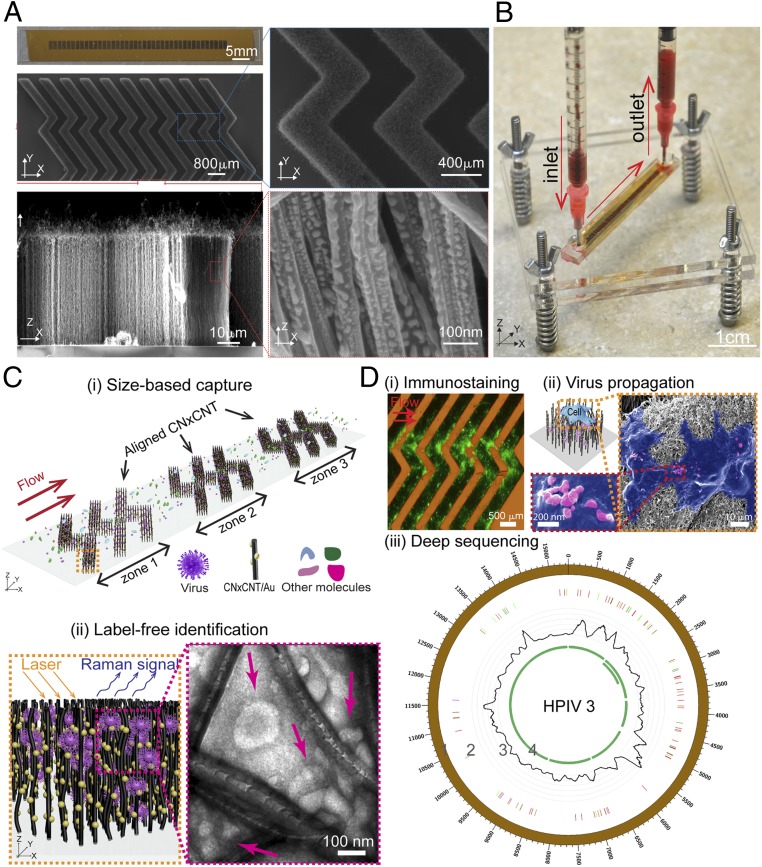
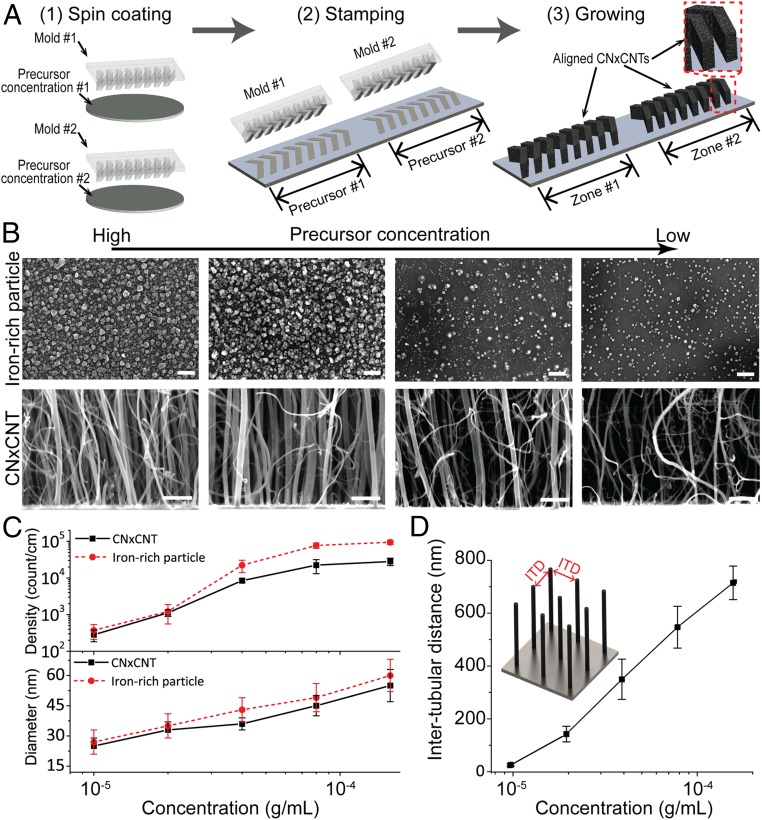
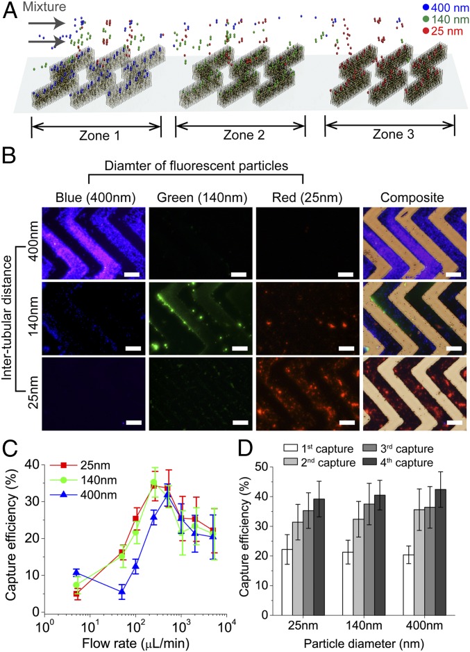
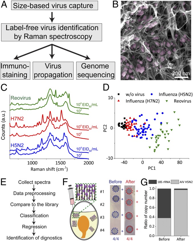
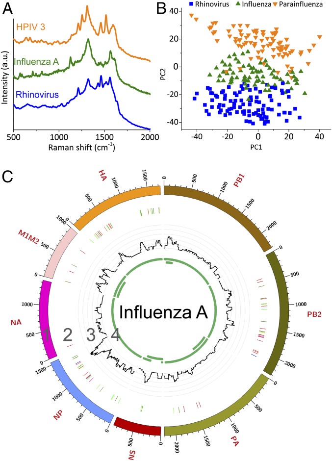
Emerging and reemerging viruses are responsible for a number of recent epidemic outbreaks. A crucial step in predicting and controlling outbreaks is the timely and accurate characterization of emerging virus strains. We present a portable microfluidic platform containing carbon nanotube arrays with differential filtration porosity for the rapid enrichment and optical identification of viruses. Different emerging strains (or unknown viruses) can be enriched and identified in real time through a multivirus capture component in conjunction with surface-enhanced Raman spectroscopy. More importantly, after viral capture and detection on a chip, viruses remain viable and get purified in a microdevice that permits subsequent in-depth characterizations by various conventional methods. We validated this platform using different subtypes of avian influenza A viruses and human samples with respiratory infections. This technology successfully enriched rhinovirus, influenza virus, and parainfluenza viruses, and maintained the stoichiometric viral proportions when the samples contained more than one type of virus, thus emulating coinfection. Viral capture and detection took only a few minutes with a 70-fold enrichment enhancement; detection could be achieved with as little as 102 EID50/mL (50% egg infective dose per microliter), with a virus specificity of 90%. After enrichment using the device, we demonstrated by sequencing that the abundance of viral-specific reads significantly increased from 4.1 to 31.8% for parainfluenza and from 0.08 to 0.44% for influenza virus. This enrichment method coupled to Raman virus identification constitutes an innovative system that could be used to quickly track and monitor viral outbreaks in real time.

Keywords: carbon nanotube; infectious disease; microfabrication; sequencing; virus isolation.


Conflict of interest statement

Competing interest statement: The authors declare no competing interest.

Figures

Fig. 1.
Fig. 1.
Design and working principle of VIRRION for effective virus capture and identification. (A) Photograph and SEM images of aligned CNTs exhibiting herringbone patterns decorated with gold nanoparticles. (B) Picture showing assembled VIRRION device, processing a blood sample. (C) Illustration of (i) size-based capture and (ii) in situ Raman spectroscopy for label-free optical virus identification. Images of electron microscopy showing captured avian influenza virus H5N2 by CNxCNT arrays. (D) On-chip virus analysis and enrichment for NGS, (i) on-chip immunostaining for captured H5N2, (ii) on-chip viral propagation through cell culture, and (iii) genomic sequencing and analysis of human parainfluenza virus type 3 (HPIV 3). Track 1: scale of the base pair position; track 2: variant analysis by mapping to strain #MF973163; color code: deletion (black), transition (A–G, fluorescent green; G–A, dark green; C–T, dark red; T–C, light red), transversion (A–C, brown; C–A, purple; A–T, dark blue; T–A, fluorescent blue; G–T, dark orange; T–G, violet; C–G, yellow; G–C, light violet); track 3: coverage; track 4: regions of open reading frames (ORFs).
Fig. 2.
Fig. 2.
Stamping technique for aligned CNxCNT growth with tunable dimensions. (A) Process flow of the stamping technique for patterning CNxCNT arrays. (B) SEM of iron-rich particles (top row) and CNxCNT (bottom row) before and after CVD synthesis. [Scale bar: 100 nm (top row); 100 nm (bottom row).] (C) Density and diameter of iron-rich particles and CNxCNTs under different precursor concentrations. (D) Tunable ITD of aligned CNxCNT array growing under different precursor concentrations.
Fig. 3.
Fig. 3.
Characterization of size-based capture. (A) Illustration of capture and separation of 3 different sizes of fluorescently labeled silica particles by VIRRION with 3 zones of ITDs. (B) Fluorescent and combined bright-field images of particles captured and separated by VIRRION into individual zones. (Scale bar: 200 µm.) (C) Capture efficiency of different silica particles captured by VIRRIONS under different flow rates. (D) Capture efficiency of silica particles after multiple repeated capture by the same VIRRION.
Fig. 4.
Fig. 4.
Characterization of avian influenza virus captured and detected by VIRRION. (A) A process flow of VIRRION for avian influenza virus surveillance and discovery. (B) SEM showing H5N2 virus particles captured by CNxCNT arrays. (C) Raman spectra of H5N2, H7N2, and reovirus collected from VIRRION. (D) Classification by PCA plot of Raman spectra collected from different avian viruses. (E) Process flow of virus identification by Raman spectroscopy with algorithm. (F) H5N2 virus propagated in ECE after viable capture and detection. (G) Ratio of copy number of H5N2 and 18S rRNA before and after VIRRION enrichment.
Fig. 5.
Fig. 5.
VIRRION testing of respiratory viruses. (A) Raman spectra. (B) PCA plot of Raman fingerprint of the different viruses. Each dot represents a collected spectrum. (C) Circos plots of coverage and variants of captured influenza viruses. Genome segment sequencing and analysis of influenza A mapped to strain A/New York/03/2016 (H3N2). Track 1: scale of the base pair position; track 2: variant analysis by mapping to strain H3N2 (KX413814–KX413821), color code: deletion (black), transition (A–G, fluorescent green; G–A, dark green; C–T, dark red; T–C, light red), transversion (A–C, brown; C–A, purple; A–T, dark blue; T–A, fluorescent blue; G–T, dark orange; T–G, violet; C–G, yellow; G–C, light violet); track 3: coverage; track 4: regions of ORF.

References

    1. Suttle C. A., Viruses in the sea. Nature 437, 356–361 (2005). - PubMed
    1. Zhang Y. Z., Wu W. C., Shi M., Holmes E. C., The diversity, evolution and origins of vertebrate RNA viruses. Curr. Opin. Virol. 31, 9–16 (2018). - PMC - PubMed
    1. Lipkin W. I., Anthony S. J., Virus hunting. Virology 479–480, 194–199 (2015). - PubMed
    1. Woolhouse M. E. J., Adair K., The diversity of human RNA viruses. Future Virol. 8, 159–171 (2013). - PMC - PubMed
    1. Peiris J. S. M., de Jong M. D., Guan Y., Avian influenza virus (H5N1): A threat to human health. Clin. Microbiol. Rev. 20, 243–267 (2007). - PMC - PubMed

Publication types

MeSH terms