. 2020 Feb 7;295(6):1704-1715.
doi: 10.1074/jbc.RA119.012053. Epub 2019 Dec 27.

Pulmonary surfactant lipids inhibit infections with the pandemic H1N1 influenza virus in several animal models

Affiliations

Pulmonary surfactant lipids inhibit infections with the pandemic H1N1 influenza virus in several animal models

Mari Numata et al. J Biol Chem. .

Abstract

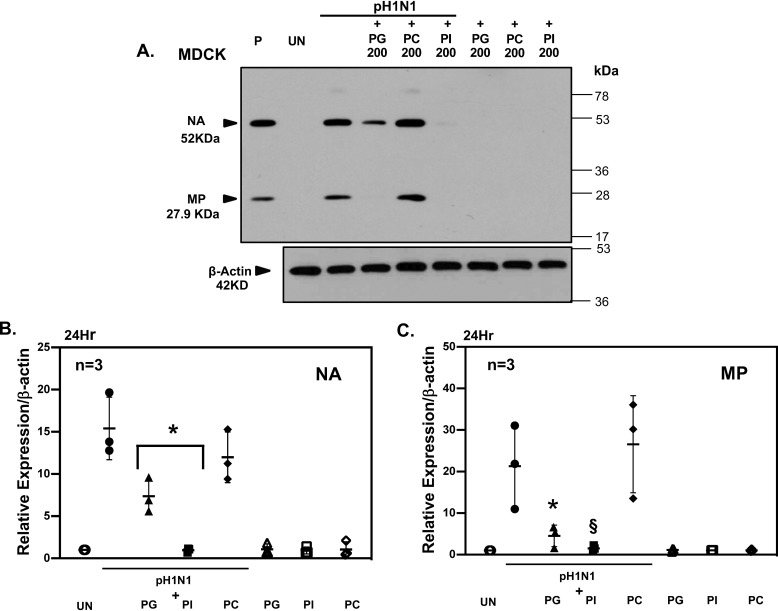
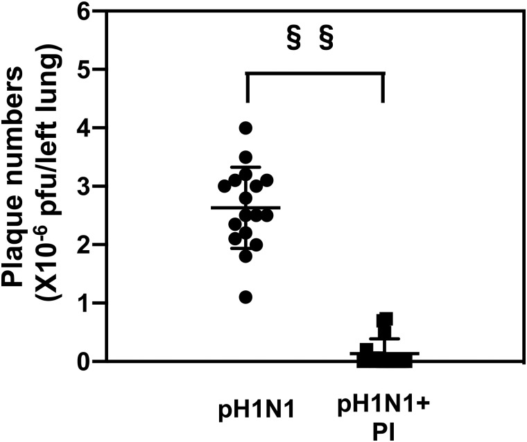
The influenza A (H1N1)pdm09 outbreak in 2009 exemplified the problems accompanying the emergence of novel influenza A virus (IAV) strains and their unanticipated virulence in populations with no pre-existing immunity. Neuraminidase inhibitors (NAIs) are currently the drugs of choice for intervention against IAV outbreaks, but there are concerns that NAI-resistant viruses can transmit to high-risk populations. These issues highlight the need for new approaches that address the annual influenza burden. In this study, we examined whether palmitoyl-oleoyl-phosphatidylglycerol (POPG) and phosphatidylinositol (PI) effectively antagonize (H1N1)pdm09 infection. POPG and PI markedly suppressed cytopathic effects and attenuated viral gene expression in (H1N1)pdm09-infected Madin-Darby canine kidney cells. POPG and PI bound to (H1N1)pdm09 with high affinity and disrupted viral spread from infected to noninfected cells in tissue culture and also reduced (H1N1)pdm09 propagation by a factor of 102 after viral infection was established in vitro In a mouse infection model of (H1N1)pdm09, POPG and PI significantly reduced lung inflammation and viral burden. Of note, when mice were challenged with a typically lethal dose of 1000 plaque-forming units of (H1N1)pdm09, survival after 10 days was 100% (14 of 14 mice) with the POPG treatment compared with 0% (0 of 14 mice) without this treatment. POPG also significantly reduced inflammatory infiltrates and the viral burden induced by (H1N1)pdm09 infection in a ferret model. These findings indicate that anionic phospholipids potently and efficiently disrupt influenza infections in animal models.

Keywords: antiviral agent; host defense; inflammation; influenza; innate immunity; phosphatidylglycerol; phosphatidylinositol; phospholipids; pulmonary surfactant; treatments; virology.


Conflict of interest statement

M. N. and D. R. V. are the co-inventors of the patent Surfactant Lipids, Compositions Thereof, and Uses Thereof; the patent is owned by National Jewish Health, Denver, CO. All of the aforementioned grants and patents are focused upon mechanisms of antiviral and anti-inflammatory actions of phospholipids

Figures

Figure 1.
Figure 1.
POPG and PI inhibit (H1N1)pdm09 protein expression and cytopathology. Cultures of MDCK cells were grown at 37 °C in DMEM plus 10% bovine growth serum with antibiotics, until reaching 100% confluence. The growth medium was removed by aspiration, and the cultures were then washed three times with PBS. The cultures were infected, at a m.o.i. = 0.5 with (H1N1)pdm09, for 24 h using growth medium without serum. Subsequently, cultures were lysed in the well by addition of 300 μl of SDS-PAGE loading buffer. The extracts were subjected to electrophoresis, and the separated proteins were transferred to nitrocellulose and probed with HRP-conjugated polyclonal antibodies recognizing viral proteins. Immunoreactive viral proteins were detected using a diaminobenzidine substrate. A, arrowheads show the migration of molecular size standards (NA and MP). The migration of additional molecular size standards (17–78 kDa) is also shown on the right side of the blot. Figure shows the migration positions of viral neuraminidase (NA) and matrix protein (MP) as well as the recovery of β-actin in the cell extracts β-actin refers to product of Actb gene. P represents viral protein detection by immunoblotting using samples from purified virus stock. UN, uninfected MDCK cell cultures; pH1N1, cells infected with (H1N1)pdm09 for 24 h at an m.o.i. = 0.5, as indicated in the figure, in either the absence or presence (+) of 200 μg of phospholipids added as liposome preparations (either PG, PC, or PI). The figure also shows the results of adding lipids in the absence of (H1N1)pdm09. B, quantitative data from three independent experiments. Experiments in A were performed three times, and the cumulative data and variation for NA are shown in B, with either uninfected (UN) cultures or those infected with (H1N1)pdm09 (pH1N1); the cumulative data for quantification of MP are shown in C. Significance: * indicates p < 0.05, and § indicates p < 0.01 when compared with pH1N1 alone. Data are means ± S.D. from three experiments.
Figure 2.
Figure 2.
(H1N1)pdm09 binds phospholipids with high affinity. Phospholipid solid phases (PG, PI, and PC) were generated by adding an ethanolic suspension of 1.5 nmol of phospholipid to microtiter wells and removing the solvent under a current of room temperature air. Subsequently, wells were washed with PBS and blocked with 3% BSA/PBS. A suspension containing purified (H1N1)pdm09 (0.03–4 × 10⁷ pfu/ml) was added to each well, and the binding reaction was performed for 1 h at 37 °C. After three washings, virus bound to the solid-phase lipid was detected using an HRP-conjugated polyclonal goat anti-influenza antibody and a diaminobenzidine substrate. Antibody binding was quantified spectrophotometrically using A450. Data are means ± S.D. for three independent experiments.
Figure 3.
Figure 3.
POPG and PI inhibit (H1N1)pdm09 attachment to MDCK cells. A, MDCK cells were grown in DMEM until confluent. The adherent cells were washed three times with PBS. Cells were either sham infected (UN) or treated with pH1N1, at a m.o.i. = 20, in either the absence or presence of 200 μg/ml phospholipids (PG, PC, and PI), as indicated. Incubations were performed for 2 h at 0 °C to minimize endocytosis. After the incubation period, wells were washed three times with PBS, and the cell layers were harvested in SDS-PAGE buffer. The cell lysates were subjected to electrophoresis, transferred to nitrocellulose, and blotted with polyclonal goat anti-IAV antibody. β-actin refers to product of Actb gene. B shows results averaged from three experiments, and data are expressed as means ± S.D. Significance: * indicates p < 0.05 when compared with virus alone.
Figure 4.
Figure 4.
POPG and PI inhibit (H1N1)pdm09 attachment to human A549 cells. A shows data for A549 cells grown, processed, and harvested as described for Fig. 3A. Cell extracts were prepared by adding SDS-PAGE loading buffer to culture wells and harvesting after repeated pipetting. The harvested cell lysates were subjected to electrophoresis, transferred to nitrocellulose, and immunoblotted with goat polyclonal anti-influenza antibodies. P indicates pattern of viral antigen separation using purified virus preparations. UN indicates immunoreactive material detected from uninfected cell layers, and pH1N1 indicates distribution of immunoreactive material detected after viral adsorption at 0 °C using (H1N1)pdm09 in either the absence or presence of phospholipids (+PG, +PI, or +PC). β-actin refers to product of Actb gene. B shows results from three independent experiments, and data are expressed as means ± S.D. §§ indicates p < 0.001, and § indicates p < 0.01, when compared with viral infection alone.
Figure 5.
Figure 5.
POPG and PI inhibit secondary (H1N1)pdm09 infection after establishment of limited primary infections in MDCK cells. Figure shows the results of representative plaque assays of (H1N1/pdm09) using simultaneous treatments with phospholipids and (H1N1)pdm09 at a concentration of 1000 μg/ml lipids (POPG, PC, and PI), and the data for plaque numbers resulting from simultaneous treatments with virus + phospholipids and post-infection treatments with phospholipids (designated pPG and pPI). Data are from three individual experiments and shown as means ± S.D. §§ indicates p < 0.001; § indicates p < 0.01; and * indicates p < 0.05.
Figure 6.
Figure 6.
POPG inhibits inflammatory cell infiltrates and IFN-γ production in response to (H1N1)pdm09 infection in mice. A, mice (5–8/group) were either sham-infected (CONL) or treated with (H1N1)pdm09 at 102 pfu/mouse, delivered intranasally (pH1N1), or virus + 3 mg of POPG (pH1N1 + PG) or only 3 mg of PG (PG), as indicated. The addition of lipids was in 50 μl of an aqueous suspension of unilamellar liposomes. The infections progressed for 6 days. B shows quantification of neutrophil populations recovered in lavage. C shows quantification of lymphocyte populations in lavage. D shows IFN-γ levels recovered in cell-free lavage. Values are means ± S.D. Significance: §§ indicates p < 0.001. Data are from three independent experiments.
Figure 7.
Figure 7.
POPG reduces the viral burden resulting from (H1N1)pdm09 infections. Mice (5–8/group) were treated with (H1N1)pdm09 at 102 pfu/mouse, delivered intranasally (pH1N1), or virus + 3 mg of POPG (pH1N1 + PG). After 6 days, mice were euthanized, and the lungs were harvested, homogenized, and then processed for viral plaque assays. Data are from three independent experiments. Values are expressed as means ± S.D. Significance: §§ indicates p < 0.001.
Figure 8.
Figure 8.
PG reduces negative histopathology associated with (H1N1)pdm09 infections. A, mice were treated with virus and lipids as described for Fig. 6. At 6 days after infection, animals were euthanized by asphyxiation with CO2, and the right lungs were harvested, fixed, sectioned, and stained with H&E solution. The sections were scored for histopathology in a blinded fashion, and the results are shown in A. B shows representative sections for uninfected (CONL), virally infected (pH1N1), virally infected and lipid-treated (pH1N1 + PG), and lipid alone (PG) treated mice. Data shown are from three independent experiments, with 5–8 mice per group in each experiment. Data are means ± S.D. Significance: §§ indicates p < 0.001 when compared with virus treatment alone.
Figure 9.
Figure 9.
PI protects mice from (H1N1)pdm09 infection. A, mice (5–8/group), were either sham-infected (CONL) or treated with (H1N1)pdm09 at 102 pfu/mouse, delivered intranasally (pH1N1), or virus + 0.6 mg of PI (pH1N1 + PI) or only 0.6 mg of PI (PI), as indicated. Lipids were added as aqueous suspensions of small unilamellar liposomes in a volume of 50 μl along with virus. B, inflammatory neutrophilic infiltrates recovered from lung lavage. C, lymphocytic infiltrates recovered from lavage after (H1N1)pdm09 infection. D, IFN-γ levels recovered from cell free lavage. Significance: §§ indicates p < 0.001; * indicates p < 0.05. Data are from three independent experiments.
Figure 10.
Figure 10.
PI reduces the viral burden elicited by (H1N1)pdm09 infection. Mice were either infected with (H1N1)pdm09, at 102 pfu/mouse (pH1N1), delivered intranasally, or infected in the presence of 0.6 mg of PI (PI). After 6 days, mice were euthanized, and the lungs were cannulated and lavaged. The right lungs were homogenized, and cell extracts were used to quantify the viral burden by plaque assays. Significance: §§ indicates p < 0.001.
Figure 11.
Figure 11.
PI attenuates the negative histopathology accompanying (H1N1)pdm09 infection. Mice were either uninfected (CONL) or infected with (H1N1)pdm09, at 102 pfu/mouse (pH1N1), delivered intranasally, or infected in the presence of 0.6 mg of PI (pH1N1 + PI), or treated with the lipid alone (PI). After 6 days, mice were euthanized, and the lungs were cannulated and lavaged, fixed, and stained with H&E. A, stained tissues were examined by light microscopy and assigned a histopathology score. B, representative lung tissue sections stained with H&E and visualized by light microscopy. Significance: §§ indicates p < 0.001.
Figure 12.
Figure 12.
PG protects mice from lethal (H1N1)pdm09 infection. A, experimental design for studying lethal (H1N1)pdm09 infection in mice. Mice were given a lethal dose of 103 pfu of (H1N1)pdm09, by intranasal inoculation, either in the absence or presence of a 3-mg dose of POPG, and then followed for 12 days. The mice were weighed each day and removed from the study and euthanized when weight loss reached 20%, or greater, of initial weight on day 0. B, survival curves following infection. Mice were removed from the study when weight loss was equal to or exceeded 20% of the starting weight. C, weight gain/loss among different experimental groups over the time course of the experiment.
Figure 13.
Figure 13.
PG ameliorates (H1N1)pdm09 infection in ferrets. Ferrets were anesthetized with isoflurane and then infected with (H1N1)pdm09 using a 500-μl inoculation containing 106 pfu of virus. After 4 days, ferrets were euthanized, and the lungs were lavaged with 30 ml of saline. A shows the total cells recovered in lavage of sham-infected (CONL) and (H1N1)pdm09-infected ferrets (pH1N1), in addition to ferrets receiving virus plus POPG (pH1N1 + PG) or lipid alone (PG). Significance: * indicates p < 0.05 when compared with virus alone. B, cell populations present in BALF were quantified after cytospin recovery, using staining, microscopy, and manual counting. Significance: * indicates p < 0.05 when comparing values between (H1N1)pdm09 (pH1N1) and (H1N1)pdm09 + PG (pH1N1 + PG).
Figure 14.
Figure 14.
PG reduces the viral burden in multiple respiratory compartments of infected ferrets. Four days following infection of ferrets as described for Fig. 13, the animals were anesthetized using isoflurane inhalation, and the nasal compartments (A) and pharynx (B) were sampled using sterile cotton swabs. After the process of sampling, the swabs were placed in sterile medium and vigorously agitated to dislodge viral particles. Ferrets were subsequently euthanized, and the lungs were lavaged with three 10-ml aliquots of isotonic saline to yield a BALF fraction (C). Recovered lavage fluid harboring viruses was measured and diluted, and infectious particles were quantified by plaque assays (A–D). Significance: * indicates p < 0.05; § indicates p < 0.01.
Figure 15.
Figure 15.
PG alleviates the negative histopathology accompanying (H1N1)pdm09 infection in ferrets. The lungs of euthanized ferrets were lavaged with three 10-ml aliquots of isotonic saline, fixed in 10% buffered formalin, and sectioned and subjected to quantitative histopathology assessment. A shows the histopathology scores of lungs from uninfected (CONL) and infected (pH1N1) ferrets, some treated with POPG (+PG) at the time of infection. Lungs were fixed with buffered formalin, and tissue sections were prepared and stained with H&E and evaluated for histopathology. Significance: * indicates p < 0.05. B shows representative sections from sham-infected (CONL) and virus-infected (pH1N1) ferrets, either untreated or treated with 5 mg of POPG (+PG). Control experiments also included treatment with lipid alone (PG). Scale bar is 100 μm, as indicated in the panels.

References

    1. Shinde V., Bridges C. B., Uyeki T. M., Shu B., Balish A., Xu X., Lindstrom S., Gubareva L. V., Deyde V., Garten R. J., Harris M., Gerber S., Vagasky S., Smith F., Pascoe N., et al. (2009) Triple-reassortant swine influenza A (H1) in humans in the United States, 2005–2009. N. Engl. J. Med. 360, 2616–2625 10.1056/NEJMoa0903812 - DOI - PubMed
    1. Garten R. J., Davis C. T., Russell C. A., Shu B., Lindstrom S., Balish A., Sessions W. M., Xu X., Skepner E., Deyde V., Okomo-Adhiambo M., Gubareva L., Barnes J., Smith C. B., Emery S. L., et al. (2009) Antigenic and genetic characteristics of swine-origin 2009 A(H1N1) influenza viruses circulating in humans. Science 325, 197–201 10.1126/science.1176225 - DOI - PMC - PubMed
    1. Centers for Disease Control and Prevention (CDC). (2012) Update: Influenza activity–United States, October 2, 2011–February 11, 2012. MMWR Morb. Mortal. Wkly. Rep. 61, 123–128 - PubMed
    1. Epperson S., Blanton L., Kniss K., Mustaquim D., Steffens C., Wallis T., Dhara R., Leon M., Perez A., Chaves S. S., Elal A. A., Gubareva L., Xu X., Villanueva J., Bresee J., et al. (2014) Influenza activity–United States, 2013–14 season and composition of the 2014–15 influenza vaccines. MMWR Morb. Mortal. Wkly. Rep. 63, 483–490 - PMC - PubMed
    1. Blanton L., Dugan V. G., Abd Elal A. I., Alabi N., Barnes J., Brammer L., Budd A. P., Burns E., Cummings C. N., Garg S., Garten R., Gubareva L., Kniss K., Kramer N., O'Halloran A., et al. (2019) Update: Influenza activity–United States, September 30, 2018-February 2, 2019. MMWR Morb. Mortal. Wkly. Rep. 68, 125–134 10.15585/mmwr.mm6806a1 - DOI - PMC - PubMed

Publication types

MeSH terms

Substances