. 2020 Feb 7;295(6):1517-1538.
doi: 10.1074/jbc.RA119.011577. Epub 2019 Dec 27.

Structural and mechanistic insights into Hsp104 function revealed by synchrotron X-ray footprinting

Affiliations

Structural and mechanistic insights into Hsp104 function revealed by synchrotron X-ray footprinting

Elizabeth A Sweeny et al. J Biol Chem. .

Abstract

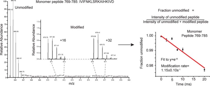
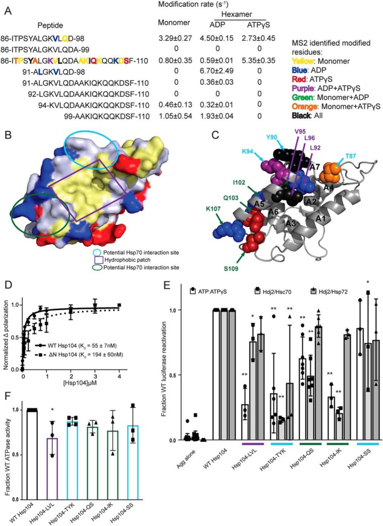
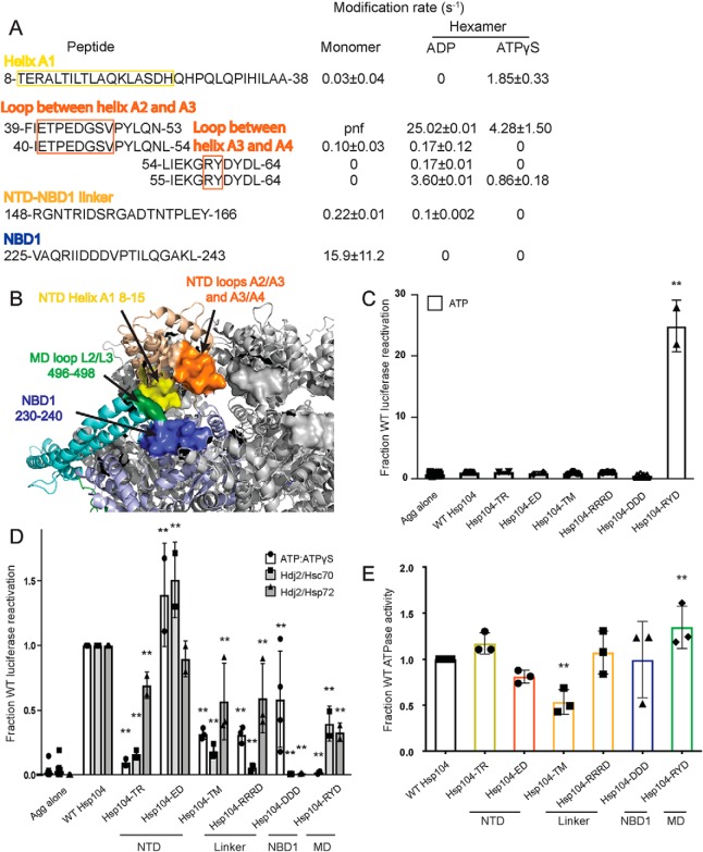
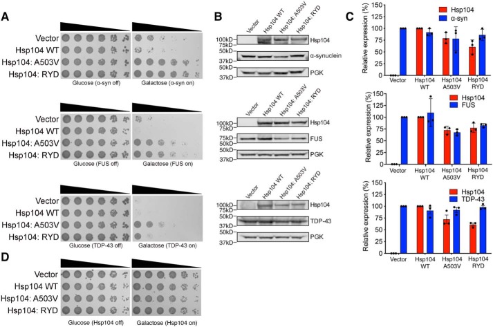
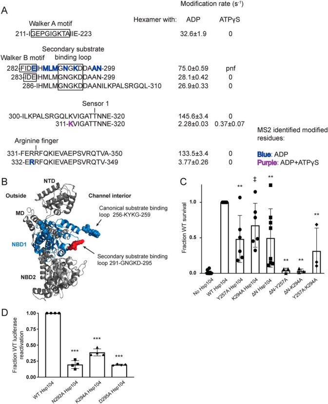
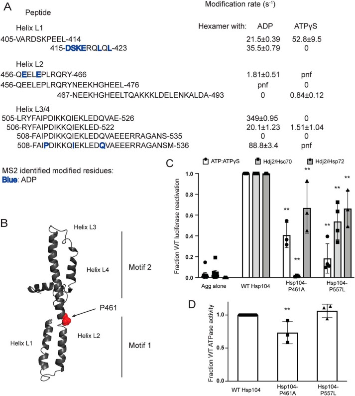
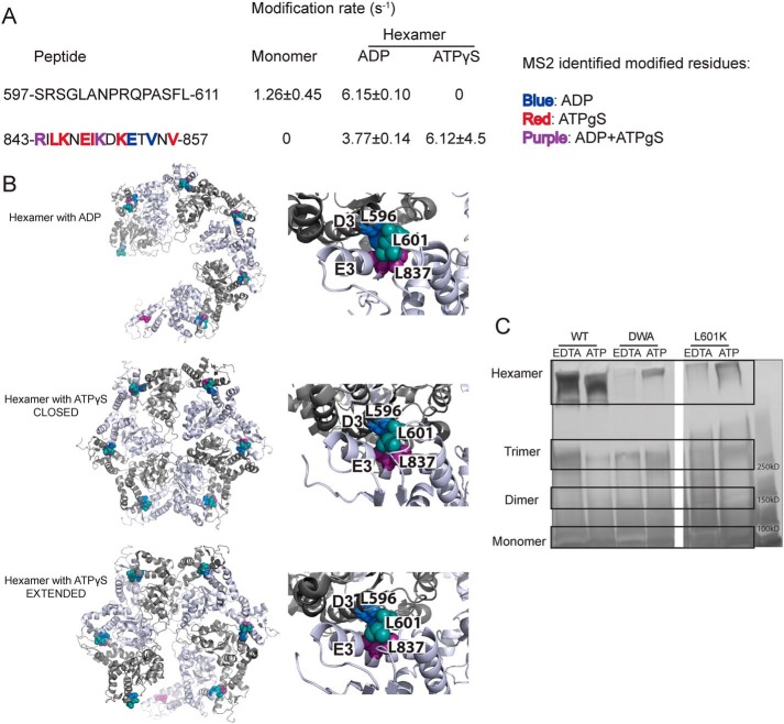
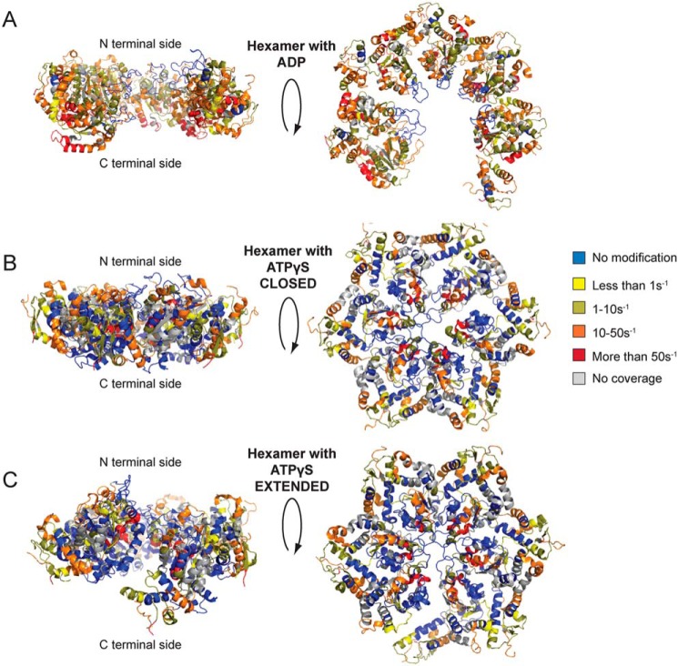
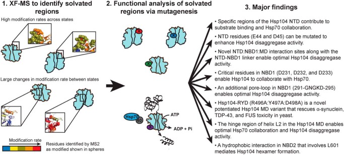
Hsp104 is a hexameric AAA+ ring translocase, which drives protein disaggregation in nonmetazoan eukaryotes. Cryo-EM structures of Hsp104 have suggested potential mechanisms of substrate translocation, but precisely how Hsp104 hexamers disaggregate proteins remains incompletely understood. Here, we employed synchrotron X-ray footprinting to probe the solution-state structures of Hsp104 monomers in the absence of nucleotide and Hsp104 hexamers in the presence of ADP or ATPγS (adenosine 5'-O-(thiotriphosphate)). Comparing side-chain solvent accessibilities between these three states illuminated aspects of Hsp104 structure and guided design of Hsp104 variants to probe the disaggregase mechanism in vitro and in vivo We established that Hsp104 hexamers switch from a more-solvated state in ADP to a less-solvated state in ATPγS, consistent with switching from an open spiral to a closed ring visualized by cryo-EM. We pinpointed critical N-terminal domain (NTD), NTD-nucleotide-binding domain 1 (NBD1) linker, NBD1, and middle domain (MD) residues that enable intrinsic disaggregase activity and Hsp70 collaboration. We uncovered NTD residues in the loop between helices A1 and A2 that can be substituted to enhance disaggregase activity. We elucidated a novel potentiated Hsp104 MD variant, Hsp104-RYD, which suppresses α-synuclein, fused in sarcoma (FUS), and TDP-43 toxicity. We disambiguated a secondary pore-loop in NBD1, which collaborates with the NTD and NBD1 tyrosine-bearing pore-loop to drive protein disaggregation. Finally, we defined Leu-601 in NBD2 as crucial for Hsp104 hexamerization. Collectively, our findings unveil new facets of Hsp104 structure and mechanism. They also connect regions undergoing large changes in solvation to functionality, which could have profound implications for protein engineering.

Keywords: ATPases associated with diverse cellular activities (AAA); FUS; Hsp104; TDP-43; disaggregase; heat-shock protein (HSP); mass spectrometry (MS); mutagenesis; protein chemical modification; α-synuclein.


Conflict of interest statement

The authors declare that they have no conflicts of interest with the contents of this article

Figures

Figure 1.
Figure 1.
X-ray exposed Hsp104 mass spectra and curve fitting. Unmodified and modified versions of the Hsp104 peptides were identified by ExMS-CL in each time point. The intensities of the singly-modified peptides were pooled, and the fraction unmodified was calculated for each time point. A pseudo–first-order decay curve extrapolated to zero was then fit to the data.
Figure 2.
Figure 2.
Hsp104 NTD is involved in substrate and Hsp70 interactions. A, peptide sequences and modification rates for a region of the Hsp104 NTD that is highly modified. Residues shown in bold have been identified as modified by MS2 and are colored by the state in which they were found: yellow, monomer; blue, hexamer with ADP; red, hexamer with ATPγS; purple, both hexameric states; green, monomer and hexamer with ADP; orange, monomer and hexamer with ATPγS; black, modified in all states. B, surface rendering of Hsp104 NTD from the crystal structure of the S. cerevisiae Hsp104 NTD (PDB code 5u2u). Acidic residues are shown in red, basic residues in blue, and hydrophobic residues in yellow. Circled regions are important in the structurally-conserved ClpA (PDB code 1r6c) NTD for ClpS binding as follows: site A in green, site C in light blue, and the conserved hydrophobic patch for substrate binding in purple. C, Hsp104 NTD with residues identified by MS2 as modified in a hexameric state are shown as spheres and labeled when visible. Spheres were colored by the states in which they were found modified, as in A, and labeled with colors corresponding to the regions outlined in B. D, FITC-casein, 2 mm ATPγS, and either Hsp104 or Hsp104ΔN were incubated for 20 min at 25 °C. Fluorescence polarization was measured at increasing concentrations of Hsp104, and a single site Kd was calculated using a least-squares fit to the data. Each data point represents mean ± S.E. (n = 4–5). E, urea-denatured firefly luciferase aggregates were incubated with Hsp104 or an Hsp104 variant, Hsp104-LVL (L92A/V95A/L96A), Hsp104-TYK (T87A/Y90A/K94A), Hsp104-QS (Q103A/S109A), Hsp104-IK (I102A/K107A), or Hsp104-SS (S124A/S125A) for 90 min at 25 °C in the presence of either 5.1 mm ATP or ATP/ATPγS; 2.6 mm ATP, and 2.5 mm ATPγS, or Hdj2/Hsc70; 5.1 mm ATP, 1 μm Hdj2, and 1 μm Hsc70, or Hdj2/Hsp72; 5.1 mm ATP, 1 μm Hdj2, and 1 μm Hsp72. Reactivation of luciferase was then determined by measuring luminescence and converted to fraction WT activity for each condition. Data are displayed as scatterplot with bar representing mean ± S.D. (n = 3–7). One-way ANOVA with Dunnett's test was performed against Hsp104-WT with * denoting p < 0.05 and ** p < 0.01. Hsp104 variants are colored based on their location as shown in B. F, ATPase activity for Hsp104 and variants. Hsp104 variants are colored based on their location in the regions highlighted in B. Data are displayed as scatterplot with bar representing mean ± S.D. (n = 3–4). One-way ANOVA with Dunnett's test comparing the variants to Hsp104-WT were performed with * denoting p < 0.05.
Figure 3.
Figure 3.
NTD–NBD1–MD interface regulates Hsp104 disaggregase activity. A and B, based on the solvation data from XF, there may be regions of the Hsp104 NTD, NBD1, and MD that make contacts that are nucleotide-dependent. In the NTD, these regions are the beginning of helix A1 in the hexamer with ADP (residues 8–15 in yellow), the A2–A3 loop (residues 39–53 in orange), and NTD–NBD1 linker (not pictured) in the hexamer with ATPγS. Based on: (1) the XF data, (2) the three monomeric tClpB crystal structures (PDB code 1qvr), and (3) the hexameric cryo-EM reconstruction (PDB code 5vy9, shown in B), we suggest that these NTD regions may be making a stable interaction with NBD1 (specifically residues 230–240 in blue) and in the hexamer with ATPγS the MD in the loop between MD helices L2 and L3 in motif 2 (residues 496–498 in green). Shifts in these contacts in response to stimuli such as Hsp70 binding, substrate binding, or ATP hydrolysis could transmit signals through the hexamer and facilitate cooperativity. In the table with rate information, pnf denotes “peptide not found.” C and D, urea-denatured firefly luciferase aggregates were incubated with either WT or an Hsp104 variant, Hsp104-TR (T8A/R10A), Hsp104-ED (E44R/D45R), Hsp104-TM (T160M), Hsp104-RRRD (R148D/R152D/R156D), Hsp104-DDD (D231A/D232A/D233A), or Hsp104–RYD (R496A/Y497A/D498A) for 90 min at 25 °C in the presence of either 5.1 mm ATP (C) or ATP/ATPγS; 2.6 mm ATP and 2.5 mm ATPγS or Hdj2/Hsc70; 5.1 mm ATP, 1 μm Hdj2, and 1 μm Hsc70 or Hdj2/Hsp72; 5.1 mm ATP, 1 μm Hdj2, and 1 μm Hsp72 (D). Reactivation of luciferase was then determined by measuring luminescence and converted to fraction WT activity for each condition. Data are displayed as scatterplot with bar representing mean ± S.D. (n = 2–4). One-way ANOVA with Dunnett's test comparing the variants to Hsp104-WT were performed with ** denoting p < 0.01. E, ATPase activity for Hsp104-WT and variants. Hsp104 variants are colored based on their location as shown in A and B. Values represent mean ± S.D. (n = 3). One-way ANOVA with Dunnett's test comparing the variants to Hsp104-WT were performed with ** denoting p < 0.01.
Figure 4.
Figure 4.
Hsp104–RYD is a potentiated variant. A, Hsp104–RYD antagonizes α-synuclein, FUS, and TDP-43 toxicity. Empty vector, Hsp104 (WT), Hsp104A503V, or Hsp104–RYD plasmids were transformed into W303aΔhsp104 yeast strains integrated with galactose-inducible α-synuclein-YFP, FUS, or TDP-43. Yeast were serially diluted 5-fold and spotted in duplicate onto galactose (inducing) and glucose (noninducing) media. B, samples were also processed for immunoblotting to assess Hsp104, PGK1 (loading control), and α-synuclein–YFP, FUS, or TDP-43 expression. Molecular mass markers are indicated (left). C, quantification of immunoblots from B. Hsp104 levels (red bars) were normalized to PGK (loading control), and the relative expression level compared with Hsp104 WT was determined. α-Synuclein–YFP, FUS, and TDP-43 levels (blue bars) were normalized to PGK (loading control), and the relative expression level compared with vector control was determined. Data are displayed as scatterplot with bar representing mean ± S.D. (n = 3). D, Hsp104–RYD is not toxic to yeast at 30 °C. Empty vectors, Hsp104 (WT), Hsp104A503V, or Hsp104–RYD plasmid were transformed into W303aΔhsp104 yeast strains. Yeast were serially diluted 5-fold and spotted in duplicate onto galactose (inducing) and glucose (noninducing) media.
Figure 5.
Figure 5.
Second substrate-binding loop in NBD1 is essential for Hsp104 disaggregase activity. A, peptide sequences and modification rates for regions of interest of Hsp104 NBD1. Residues shown in bold have been identified as modified by MS2 and are colored by the state in which they were found: blue, hexamer with ADP; purple, both hexameric states. B, NBD1 homology modeled on the tClpB crystal structure (PDB code 1qvr) shown in context of a rigid body fit Hsp104 monomer. NBD1 is shown in blue, with the canonical substrate-binding loop in blue spheres and the secondary substrate-binding loop in red spheres. C, after incubation at 37 °C for 30 min to induce Hsp104 expression, W303aΔhsp104 yeast carrying either empty vector or a plasmid encoding the indicated Hsp104 variant were heat-shocked for 20 min at 50 °C, immediately transferred to ice for 2 min, plated on SD-ura plates, and after a 2-day incubation at 30 °C, colonies were counted using an acolyte automated colony counter. Data are displayed as scatterplot with bar representing mean ± S.D. (n = 3–14). Equal expression levels were confirmed by immunoblot. A one-way ANOVA with Dunnett's test was used to compare WT Hsp104 to the variants with ‡ denoting p = 0.05 and ** denoting p < 0.01. D, urea-denatured firefly luciferase aggregates were incubated with either WT or an Hsp104 substrate-binding loop 2 (291GNGKD295) variant, for 90 min at 25 °C in the presence of 2.6 mm ATP, 2.5 mm ATPγS, and an ATP-regenerating system (1 mm creatine phosphate and 0.25 μm creatine kinase). Reactivation of luciferase was then determined by measuring luminescence and converted to fraction WT activity for each condition. Data are displayed as scatterplot with bar representing mean ± S.D. (n = 3). A one-way ANOVA with Dunnett's test comparing variants to Hsp104-WT was performed with *** denoting p < 0.0001.
Figure 6.
Figure 6.
Hsp104 MD hinge region is crucial for Hsp70 collaboration and intrinsic Hsp104 disaggregase activity. A, peptide sequences and modification rates for regions of interest of Hsp104 MD. Residues shown in bold have been identified as modified by MS2 and are colored by the state in which they were found: blue, hexamer with ADP. B, isolated MD modeled off the tClpB crystal structure (PDB code 1qvr). Helices L1–L4 are labeled, and residue Pro-461 is shown as spheres. C, urea-denatured firefly luciferase aggregates were incubated with either WT or an Hsp104 variant, Hsp104-P461A or Hsp104-P557L for 90 min at 25 °C in the presence of either 5.1 mm ATP or ATP/ATPγS; 2.6 mm ATP and 2.5 mm ATPγS, or Hdj2/Hsc70; 5.1 mm ATP, 1 μm Hdj2, and 1 μm Hsc70, or Hdj2/Hsp72; 5.1 mm ATP, 1 μm Hdj2, and 1 μm Hsp72. Reactivation of luciferase was then determined by measuring luminescence and converted to fraction WT activity for each condition. Data are displayed as scatterplot with bar representing mean ± S.D. (n = 3). One-way ANOVA with Dunnett's test comparing the variants to Hsp104-WT were performed with ** denoting p < 0.01. D, ATPase activity for Hsp104-WT and variants. Data are displayed as scatterplot with bar representing mean ± S.D. (n = 3). One-way ANOVA with Dunnett's test comparing the variants to Hsp104-WT were performed with ** denoting p < 0.01.
Figure 7.
Figure 7.
Leu-601 plays an important role in Hsp104 hexamerization. A, peptide sequences and modification rates for regions of interest of Hsp104 NBD2. Residues shown in bold have been identified as modified by MS2 and are colored by the state in which they were found: blue, hexamer with ADP; red, hexamer with ATPγS; purple, both hexameric states. B, hexameric models (hexamer with ADP, PDB code 5vy8; hexamer with ATPγS closed, PDB code 5vjh, and extended, PDB code 5vya, conformations) of NBD2 generated from cryo-EM studies (16) showing the location of hydrophobic residues found on helices D3 and E3. On the right-hand side are higher magnification views of the interface between residues 586AIKAVSNAVRLSRSGL601 of the large subdomain of subunit 1 (helix D3) and residues 836ILNKLALRILKNEI849 of the small domain of subunit 2 (helix E3). Residues Leu-601 (large domain D3, teal), Leu-596 (large domain D3, blue), and Leu-837 (small domain E3, purple) are shown as spheres. C, glutaraldehyde cross-linking. Hsp104 shows a robust ability to form hexamers even in the presence of EDTA and absence of ATP. The double Walker A (DWA) mutant displays defects in hexamerization even in the presence of ATP. The mutant L601K, which resides in the proposed hexamer interface but outside of any secondary structure, also displays defects in the ability to form hexamers.
Figure 8.
Figure 8.
Hsp104 NBD2 cryo-EM models colored based on XF modification rates. A–C, hexameric NBD2 domains were modeled based on cryo-EM studies (16) and colored based on the XF modification rates with blue as unmodified, yellow less than 1 s−1, dark yellow 1–10 s−1, orange 10–50 s−1, red more than 50 s−1, and gray is no coverage for the hexamer with ADP (A, PDB code 5vy8) and ATPγS in the closed (B, PDB code 5vjh) and extended conformations (C, PDB code 5vya).
Figure 9.
Figure 9.
Summary of methodology and major findings. Hsp104 monomer and hexamer with ADP or ATPγS were subjected to increasing exposure times of synchrotron-generated X-rays. Mass spectrometer analysis of the samples yielded oxidative modification rates for pepsin-derived peptides, and MS2 data identified specific sites of modification. Using the modification rates as a proxy for solvation, Hsp104 variants were designed with substitutions in residues found to be highly solvated or in regions that displayed large changes in solvation across the different states. Activity of these Hsp104 variants was then assessed using in vitro and in vivo assays to determine their role in Hsp104 function.

References

    1. Shorter J. (2008) Hsp104: a weapon to combat diverse neurodegenerative disorders. Neurosignals 16, 63–74 10.1159/000109760 - DOI - PubMed
    1. Jackrel M. E., and Shorter J. (2017) Protein-remodeling factors as potential therapeutics for neurodegenerative disease. Front. Neurosci. 11, 99 10.3389/fncom.2017.00099 - DOI - PMC - PubMed
    1. Mack K. L., and Shorter J. (2016) Engineering and evolution of molecular chaperones and protein disaggregases with enhanced activity. Front. Mol. Biosci. 3, 8 10.3389/fmolb.2016.00008 - DOI - PMC - PubMed
    1. Shorter J. (2016) Engineering therapeutic protein disaggregases. Mol. Biol. Cell 27, 1556–1560 10.1091/mbc.E15-10-0693 - DOI - PMC - PubMed
    1. Shorter J. (2017) Designer protein disaggregases to counter neurodegenerative disease. Curr. Opin. Genet. Dev. 44, 1–8 10.1016/j.gde.2017.01.008 - DOI - PMC - PubMed

Publication types

MeSH terms

Substances

LinkOut - more resources