Review
. 2020 Apr;69(4):550-558.
doi: 10.2337/dbi19-0002. Epub 2019 Dec 27.

The Local Paracrine Actions of the Pancreatic α-Cell

Affiliations
Review

The Local Paracrine Actions of the Pancreatic α-Cell

Rayner Rodriguez-Diaz et al. Diabetes. 2020 Apr.

Abstract

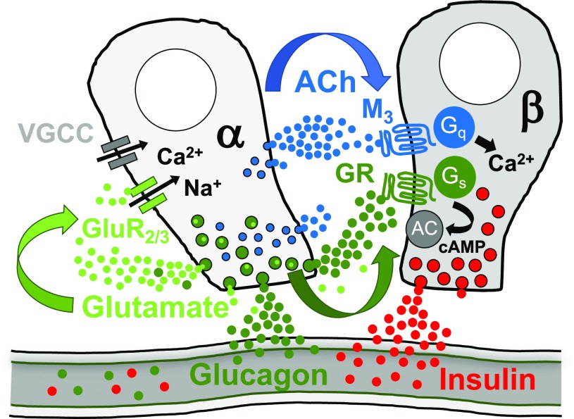
Secretion of glucagon from the pancreatic α-cells is conventionally seen as the first and most important defense against hypoglycemia. Recent findings, however, show that α-cell signals stimulate insulin secretion from the neighboring β-cell. This article focuses on these seemingly counterintuitive local actions of α-cells and describes how they impact islet biology and glucose metabolism. It is mostly based on studies published in the last decade on the physiology of α-cells in human islets and incorporates results from rodents where appropriate. As this and the accompanying articles show, the emerging picture of α-cell function is one of increased complexity that needs to be considered when developing new therapies aimed at promoting islet function in the context of diabetes.


Figures

Figure 1
Figure 1
Detection of acetylcholine (ACh) secretion from human islets using high-performance liquid chromatography and biosensor cells. A: Chromatogram showing acetylcholine detected from a 20-μL human islet sample. The acetylcholine peak area corresponds to ∼1–3 nmol/L and is accompanied by a larger choline peak. A blow up is shown on the right. Human islets were homogenized, protein was denatured, spun down, and placed in high-performance liquid chromatography column. Data contributed by Dr. Ed Phelps, University of Florida. B: Confocal images of vAChT (green) and glucagon (red) immunostaining in pancreas section from a monkey (Macaca fascicularis). As in the human, α-cells are labeled for this cholinergic marker. Panels on the right are higher magnifications of those on the left; bottom panels are merged images of vAChT and glucagon immunostaining. Scale bars = 100 μm (left), 20 μm (right). C: Trace of Ca2+ responses in biosensor cells expressing the muscarinic receptor M3 to monitor acetylcholine secretion from monkey islets (M. fascicularis). Reducing the glucose concentration from 11 mmol/L to 1 mmol/L elicited strong acetylcholine secretion. The dotted line denotes time of changes in glucose concentration. For these experiments, monkey islets were placed on a carpet of biosensor cells (trace shows mean ± SEM of 15 biosensor cells).
Figure 2
Figure 2
Cholinergic innervation of pancreatic polypeptide (PP)-secreting islet γ-cells. A: Maximum projection of a z-stack of confocal images showing cholinergic nerves labeled for vAChT (green) in a human pancreas section. Note that the density of nerves is low in the islet region demarcated by α-cells (glucagon [cyan]). By contrast, many cholinergic axons can be seen in pancreatic polypeptide–rich regions (red [at low and high magnification in A and A’]). B: As in A, cholinergic axons can be seen closely apposed to pancreatic polypeptide–labeled cells. Shown is a rare islet mainly composed of pancreatic polypeptide cells from the posterior part of the human pancreatic head. Scale bars: 50 μm (A and B), 10 μm (A’ and B’).
Figure 3
Figure 3
Cholinergic markers in the human pancreatic islet. A: Confocal images showing immunostaining for the acetylcholine-synthesizing enzyme ChAT (green [shown alone in left panel]) in human pancreas sections using tyramide signal amplification. Glucagon staining is shown in middle panel (red). Most of the ChAT staining colocalizes with glucagon (merged image in right panel). Note that some ChAT-labeled axons can be seen in the exocrine regions. B: Brightfield images showing immunostaining for ChAT using the avidin-biotin complex method. Note that a subset of islet cells is stained in the islet. Right panel is a magnification of left panel. C: Confocal images showing staining for the axonal marker synapsin (green) and the cholinergic marker vAChT (red). An axon close to the islet is labeled for both markers (arrow [staining appears yellow in the merged image on the left]). By contrast, an axon penetrating the islet is only labeled for synapsin (*). Scale bars: 100 μm (A and B), 10 μm (C).
Figure 4
Figure 4
Schematic for paracrine signaling originating from human α-cells. α-Cells secrete glucagon, acetylcholine, and glutamate, which all have been shown to have local paracrine or autocrine excitatory effects. Not depicted are additional receptors or non–β-cell targets for these signaling molecules within the islet (for more information, see acetylcholine: a shifting story). AC, adenylate cyclase; ACh, acetylcholine; GluR2/3, glutamate AMPA receptor 2/3; Gq, Gq alpha unit; GR, glucagon receptor; Gs, Gs alpha unit, G stimulatory; M3, acetylcholine muscarinic receptor 3; VGCC, voltage-gated Ca2+ channel.

References

    1. Ricordi C, Lacy PE, Finke EH, Olack BJ, Scharp DW. Automated method for isolation of human pancreatic islets. Diabetes 1988;37:413–420 - PubMed
    1. Kilimnik G, Jo J, Periwal V, Zielinski MC, Hara M. Quantification of islet size and architecture. Islets 2012;4:167–172 - PMC - PubMed
    1. Kim A, Miller K, Jo J, Kilimnik G, Wojcik P, Hara M. Islet architecture: a comparative study. Islets 2009;1:129–136 - PMC - PubMed
    1. Cabrera O, Berman DM, Kenyon NS, Ricordi C, Berggren PO, Caicedo A. The unique cytoarchitecture of human pancreatic islets has implications for islet cell function. Proc Natl Acad Sci U S A 2006;103:2334–2339 - PMC - PubMed
    1. Brissova M, Fowler MJ, Nicholson WE, et al. . Assessment of human pancreatic islet architecture and composition by laser scanning confocal microscopy. J Histochem Cytochem 2005;53:1087–1097 - PubMed

Publication types