

doi: 10.1038/s41598-019-56395-x.
Differentiation of thyroid nodules on US using features learned and extracted from various convolutional neural networks
Affiliations
- PMID: 31882683
- PMCID: PMC6934479
- DOI: 10.1038/s41598-019-56395-x
Item in Clipboard
Differentiation of thyroid nodules on US using features learned and extracted from various convolutional neural networks
Sci Rep.
.
Abstract
Thyroid nodules are a common clinical problem. Ultrasonography (US) is the main tool used to sensitively diagnose thyroid cancer. Although US is non-invasive and can accurately differentiate benign and malignant thyroid nodules, it is subjective and its results inevitably lack reproducibility. Therefore, to provide objective and reliable information for US assessment, we developed a CADx system that utilizes convolutional neural networks and the machine learning technique. The diagnostic performances of 6 radiologists and 3 representative results obtained from the proposed CADx system were compared and analyzed.
Conflict of interest statement
The authors declare no competing interests.
Figures
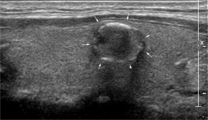
An ultrasonography (US) image of a 50-year-old woman with an incidentally detected thyroid nodule discovered on screening examination that shows a 1.2-cm sized hypoechoic solid nodule with eggshell calcifications (arrows). All 6 radiologists interpreted the nodule as a benign. In contrast, 3 CNN-combinations interpreted it as cancer. The nodule was diagnosed as papillary thyroid cancer by surgery.
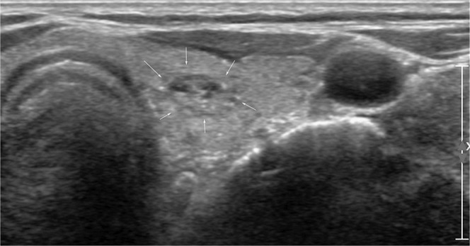
An ultrasonography (US) image of a left thyroid nodule in a 77-year-old woman who was confirmed with cancer in the right thyroid gland. A 1-cm sized isoechoic nodule with internal echogenic spots was seen (arrows). Four radiologists (1 faculty, 1 fellow, and two residents) interpreted the nodule as cancer. In contrast, 3 CNN-combinations interpreted it as benign. The nodule was diagnosed as adenomatous hyperplasia.
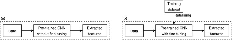
Two feature extraction strategies using pre-trained CNN: Feature extraction from pre-trained CNN without fine-tuning (a) or with fine-tuning (b).
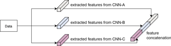
Example of feature concatenation: Feature concatenation of features extracted from three different CNNs.
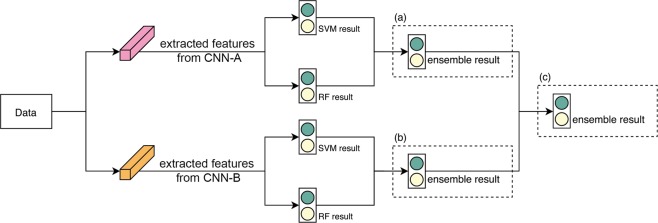
Example of classification ensemble: Two CNNs were used as feature extractors and then classification ensembles were applied for SVM and RF of CNN-A(a) and CNN-B(b) to observe results. For further objective results, the classification ensemble was again applied for ensemble results(c).
References
-
- Guth, S., Theune, U., Aberle, J., Galach, A. & Bamberger, C. J. E. J. O. C. I. Very high prevalence of thyroid nodules detected by high frequency (13 MHz) ultrasound examination. 39, 699–706 (2009). - PubMed
-
- Lim, K. J. et al. Computer-aided diagnosis for the differentiation of malignant from benign thyroid nodules on ultrasonography. 15, 853–858 (2008). - PubMed
Publication types
MeSH terms
LinkOut - more resources
Full Text Sources
