. 2019 Dec 27;9(1):19850.
doi: 10.1038/s41598-019-56427-6.

Feasibility of improving vascular imaging in the presence of metallic stents using spectral photon counting CT and K-edge imaging

Affiliations

Feasibility of improving vascular imaging in the presence of metallic stents using spectral photon counting CT and K-edge imaging

Monica Sigovan et al. Sci Rep. .

Abstract

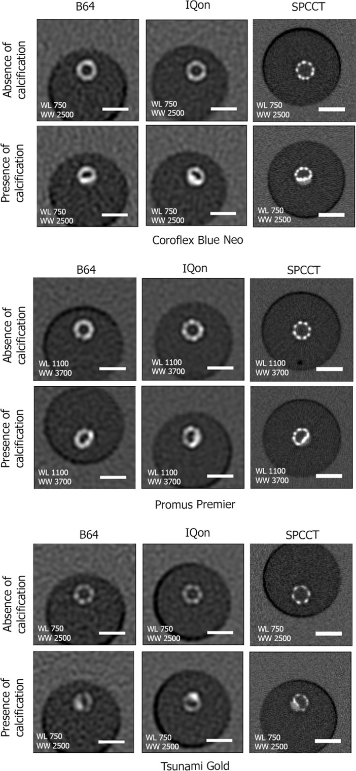
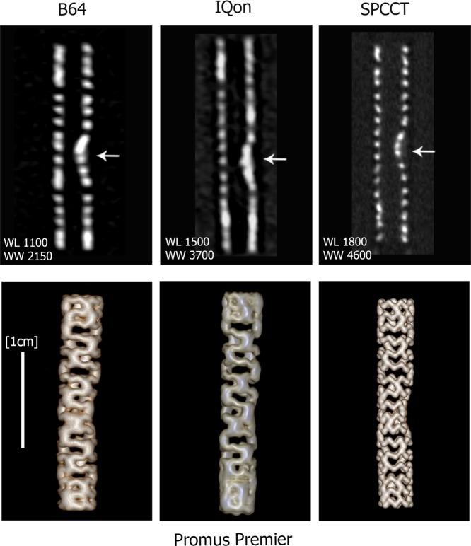
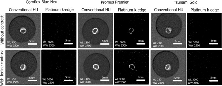
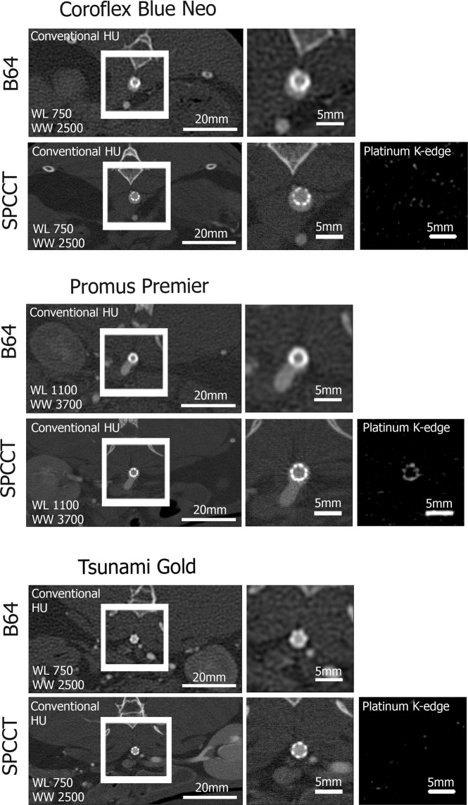
Correct visualization of the vascular lumen is impaired in standard computed tomography (CT) because of blooming artifacts, increase of apparent size, induced by metallic stents and vascular calcifications. Recently, due to the introduction of photon-counting detectors in the X-ray imaging field, a new prototype spectral photon-counting CT (SPCCT) based on a modified clinical CT system has been tested in a feasibility study for improving vascular lumen delineation and visualization of coronary stent architecture. Coronary stents of different metal composition were deployed inside plastic tubes containing hydroxyapatite spheres to simulate vascular calcifications and in the abdominal aorta of one New Zealand White (NZW) rabbit. Imaging was performed with an SPCCT prototype, a dual-energy CT system, and a conventional 64-channel CT system (B64). We found the apparent widths of the stents significantly smaller on SPCCT than on the other two systems in vitro (p < 0.01), thus closer to the true size. Consequently, the intra-stent lumen was significantly larger on SPCCT (p < 0.01). In conclusion, owing to the increased spatial resolution of SPCCT, improved lumen visualization and delineation of stent metallic mesh is possible compared to dual-energy and conventional CT.


Conflict of interest statement

P.C., I.B., M.R. and E.R. are employees of Philips Healthcare. S.S.M., M.S., P.D., L.B., J.B.L., J.M., D.B. and G.R. declare no potential conflict of interest

Figures

Figure 1
Figure 1
Representative conventional HU images acquired on the B64, the IQon, and the SPCCT of the three stents in vitro in water. Transverse views of the stents at similar locations in the absence (first row) and presence of calcification (second row). The better spatial resolution of the SPPCT system results in an improved visualization of the stent metallic struts. On the SPCCT images the stents can be visually separated from the calcification, while this separation is not possible on the B64 and IQon images due to larger detector size. A 5 mm scale bar is shown on the transverse views.
Figure 2
Figure 2
Longitudinal views of the entire length of the platinum coated stent (Promus Premier) are presented in the top row and 3D Volume Rendering are presented in the bottom row. The improved quality of SPCCT images allows clear visualization of the metallic mesh of the stent and its deformation due to the presence of the calcification insert (arrows indicate the calcification), while this is not possible for IQon and B64.
Figure 3
Figure 3
Representative conventional HU images and Pt-specific K-edge images acquired on the SPCCT in the calcification containing regions of the three different stents. The platinum coated Promus Premier stent is clearly visible on the Pt specific K-edge images while all other attenuation sources such as the calcification and the intra-luminal iodinated contrast agent are absent from these images. The clear visualization of the deformation of the metallic mesh due to the calcification is to be noted. A 5 mm scale bar is shown on each image.
Figure 4
Figure 4
Representative in vivo images of the three coronary stents placed in the abdominal aorta of a NZW rabbit. Conventional HU images are presented on the two left columns of the figure. Importantly, the Pt-specific K-edge images of SPCCT (right column) enable visualization of the Pt coated stent while all other sources of attenuation are removed.

References

    1. Dehmer GJ, et al. A contemporary view of diagnostic cardiac catheterization and percutaneous coronary intervention in the United States: a report from the CathPCI Registry of the National Cardiovascular Data Registry, 2010 through June 2011. J. Am. Coll. Cardiol. 2012;60:2017–2031. doi: 10.1016/j.jacc.2012.08.966. - DOI - PubMed
    1. Gilard M, et al. Assessment of coronary artery stents by 16 slice computed tomography. Heart Br. Card. Soc. 2006;92:58–61. doi: 10.1136/hrt.2004.056010. - DOI - PMC - PubMed
    1. Taguchi K, Iwanczyk JS. Vision 20/20: Single photon counting x-ray detectors in medical imaging. Med. Phys. 2013;40:100901. doi: 10.1118/1.4820371. - DOI - PMC - PubMed
    1. Leng S, et al. Dose-efficient ultrahigh-resolution scan mode using a photon counting detector computed tomography system. J. Med. Imaging Bellingham Wash. 2016;3:043504. doi: 10.1117/1.JMI.3.4.043504. - DOI - PMC - PubMed
    1. Symons R, et al. Photon-Counting Computed Tomography for Vascular Imaging of the Head and Neck: First In Vivo Human Results. Invest. Radiol. 2018;53:135–142. doi: 10.1097/RLI.0000000000000418. - DOI - PMC - PubMed

Publication types