. 2019 Dec 27;9(1):19887.
doi: 10.1038/s41598-019-56220-5.

Acute pancreatitis promotes the generation of two different exosome populations

Affiliations

Acute pancreatitis promotes the generation of two different exosome populations

A Jiménez-Alesanco et al. Sci Rep. .

Abstract

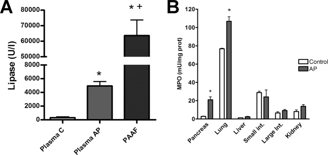
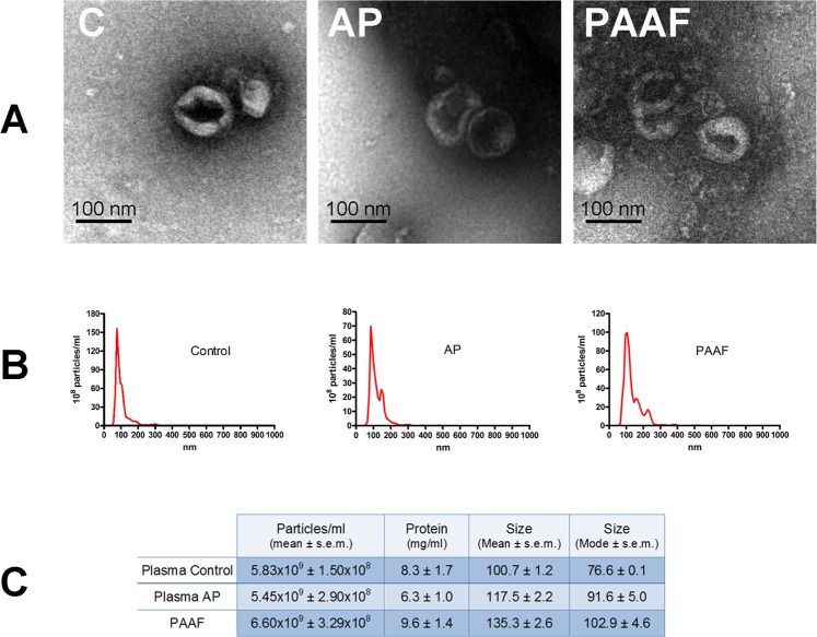
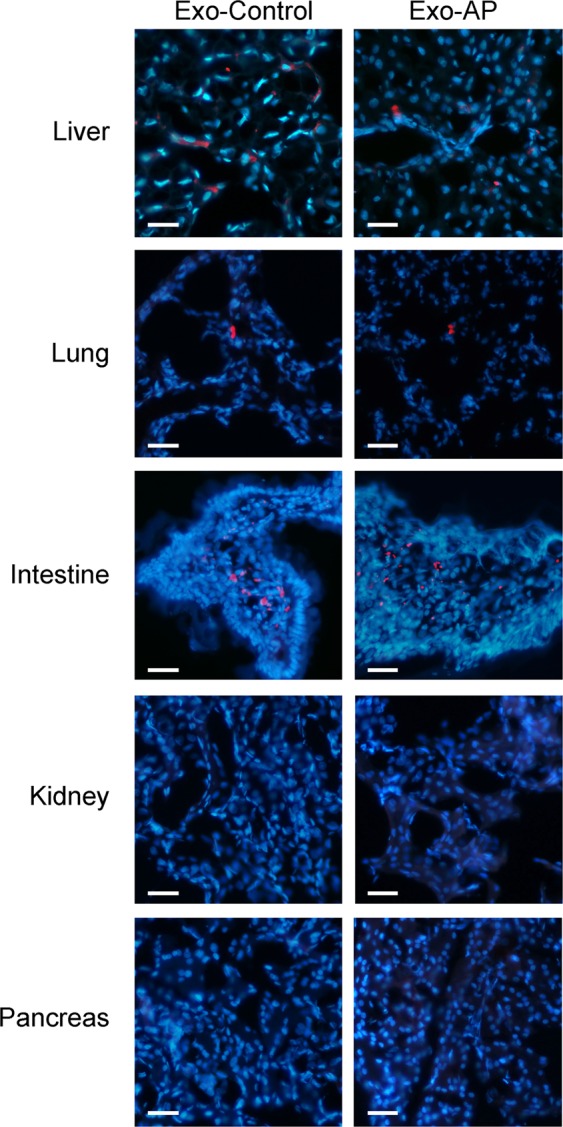
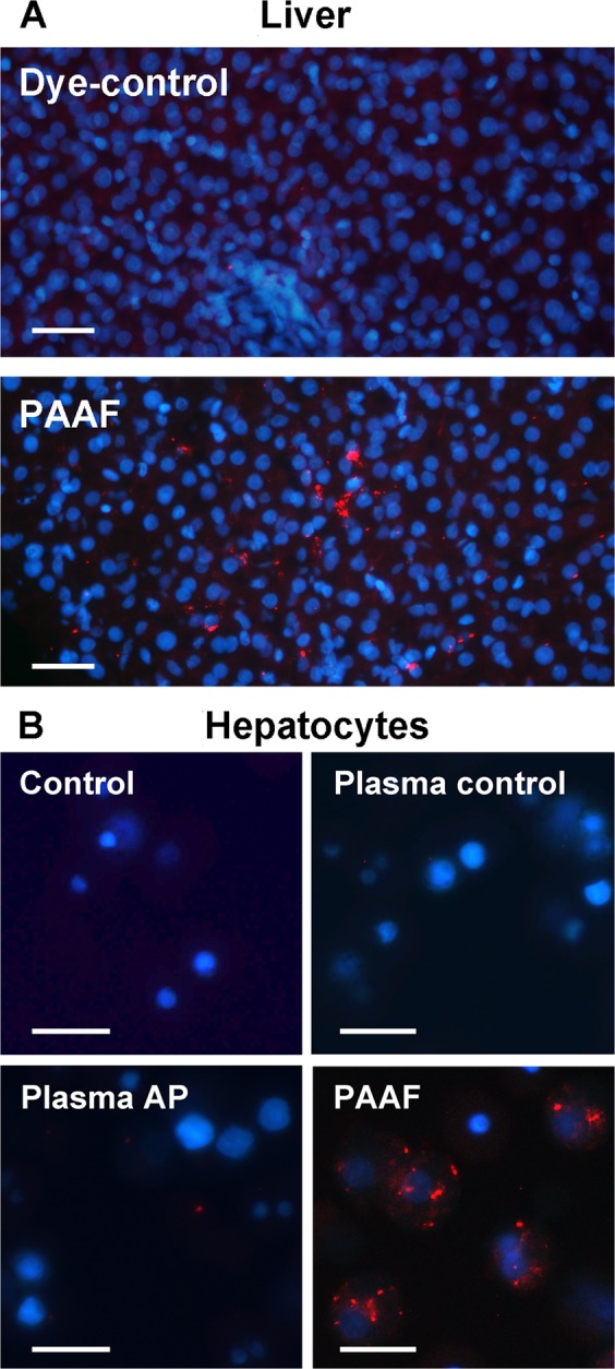
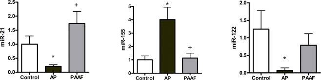
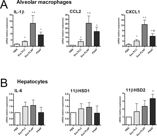
Exosomes are small extracellular vesicles that act as intercellular messengers. Previous studies revealed that, during acute pancreatitis, circulating exosomes could reach the alveolar compartment and activate macrophages. However, proteomic analysis suggested that the most likely origin of these exosomes could be the liver instead of the pancreas. The present study aimed to characterize the exosomes released by pancreas to pancreatitis-associated ascitic fluid (PAAF) as well as those circulating in plasma in an experimental model of taurocholate-induced acute pancreatitis in rats. We provide evidence that during acute pancreatitis two different populations of exosomes are generated with relevant differences in cell distribution, protein and microRNA content as well as different implications in their physiological effects. During pancreatitis plasma exosomes, but not PAAF exosomes, are enriched in the inflammatory miR-155 and show low levels of miR-21 and miR-122. Mass spectrometry-based proteomic analysis showed that PAAF exosomes contains 10-30 fold higher loading of histones and ribosomal proteins compared to plasma exosomes. Finally, plasma exosomes have higher pro-inflammatory activity on macrophages than PAAF exosomes. These results confirm the generation of two different populations of exosomes during acute pancreatitis. Deep understanding of their specific functions will be necessary to use them as therapeutic targets at different stages of the disease.


Conflict of interest statement

The authors declare no competing interests.

Figures

Figure 1
Figure 1
(A) Lipase in plasma from control (C) or acute pancreatitis (AP) animals and ascitic fluid (PAAF) and (B) myeloperoxidase (MPO) activity in different tissues 3 h after induction of acute pancreatitis. *p < 0.05 vs control;+p < 0.05 vs AP.
Figure 2
Figure 2
(A) Representative electron microscopy images of exosomes purified from plasma control (C), plasma from pancreatitis (AP) and Pancreatitis associated ascitic fluid (PAAF). (B) Nanovesicle tracking assay of extracellular vesicles. (C) Total number, protein concentration and size of particles obtained in plasma control, pancreatitis and PAAF.
Figure 3
Figure 3
Exosomes obtained from Control and AP plasma samples were stained with PKH-26 dye (red) and perfused through the inferior vena cava. Tissue samples were obtained 30 min after administration. Nuclei were stained with DAPI (blue) (Scale bar: 50 µm).
Figure 4
Figure 4
(A) Exosomes from PAAF were stained with PKH-26 dye (red) and perfused through the portal vein. PBS stained with PKH26 has been infused as a Dye-control group. Samples were obtained 30 min after administration. Nuclei were stained with DAPI (blue) (Scale: 50 µm). (B) Isolated hepatocytes were incubated in vitro for 30 min. with exosomes (red) from plasma control, plasma AP or PAAF. Control group was incubated with PBS stained with PKH26 (Scale: 20 µm).
Figure 5
Figure 5
After induction of acute pancreatitis, exosomes from plasma transport higher levels of miR-155 and lower levels of miR-21 and miR-122. By contrast, no differences in those miRNAs were observed when comparing exosomes from PAAF and from plasma control. *p < 0.05 vs control; +p < 0.05 vs AP.
Figure 6
Figure 6
(A) Effect of exosomes (2 µg/ml) in macrophage activation. Even the exosomes from plasma control (exo PLC) results in a small activation of macrophages. However, exosomes from plasma AP (exo PLAP) induces higher induction of IL1β, CCL2 and CXCL1. PAAF exosomes also increases the expression of these mediators but not achieves statistical significance compared to control plasma. Control cells are treated with the equivalent volume of PBS. (B) In hepatocytes the only effect observed was an increase in the expression of 11βHSD2 under the treatment with PAAF exosomes. *p < 0.05 vs control; +p < 0.05 vs Exo PLC; #p < 0.05 vs Exo PLAP.

References

    1. Peery AF, et al. Burden and Cost of Gastrointestinal, Liver, and Pancreatic Diseases in the United States: Update 2018. Gastroenterology. 2019;156:254–272.e11. doi: 10.1053/j.gastro.2018.08.063. - DOI - PMC - PubMed
    1. Banks PA, et al. Classification of acute pancreatitis–2012: revision of the Atlanta classification and definitions by international consensus. Gut. 2013;62:102–11. doi: 10.1136/gutjnl-2012-302779. - DOI - PubMed
    1. Singh VK, Moran RA, Afghani E, de-Madaria E. Treating acute pancreatitis: what’s new? Expert Rev. Gastroenterol. Hepatol. 2015;9:901–11. doi: 10.1586/17474124.2015.1048225. - DOI - PubMed
    1. Hirota M, et al. Relationship between plasma cytokine concentration and multiple organ failure in patients with acute pancreatitis. Pancreas. 2000;21:141–6. doi: 10.1097/00006676-200008000-00006. - DOI - PubMed
    1. Gutierrez PT, Folch-Puy E, Bulbena O, Closa D. Oxidised lipids present in ascitic fluid interfere with the regulation of the macrophages during acute pancreatitis, promoting an exacerbation of the inflammatory response. Gut. 2008;57:642–8. doi: 10.1136/gut.2007.127472. - DOI - PubMed

Publication types