. 2019 Dec 27;9(1):20199.
doi: 10.1038/s41598-019-56481-0.

In Vivo Activity of Amodiaquine against Ebola Virus Infection

Affiliations

In Vivo Activity of Amodiaquine against Ebola Virus Infection

Lisa Evans DeWald et al. Sci Rep. .

Abstract

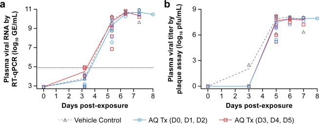
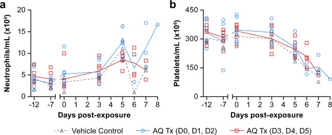
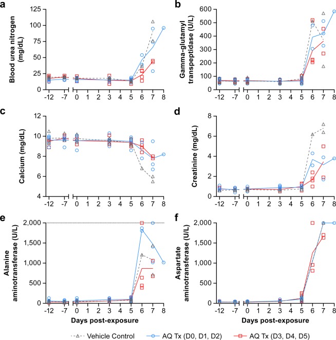
During the Ebola virus disease (EVD) epidemic in Western Africa (2013‒2016), antimalarial treatment was administered to EVD patients due to the high coexisting malaria burden in accordance with World Health Organization guidelines. In an Ebola treatment center in Liberia, EVD patients receiving the combination antimalarial artesunate-amodiaquine had a lower risk of death compared to those treated with artemether-lumefantrine. As artemether and artesunate are derivatives of artemisinin, the beneficial anti-Ebola virus (EBOV) effect observed could possibly be attributed to the change from lumefantrine to amodiaquine. Amodiaquine is a widely used antimalarial in the countries that experience outbreaks of EVD and, therefore, holds promise as an approved drug that could be repurposed for treating EBOV infections. We investigated the potential anti-EBOV effect of amodiaquine in a well-characterized nonhuman primate model of EVD. Using a similar 3-day antimalarial dosing strategy as for human patients, plasma concentrations of amodiaquine in healthy animals were similar to those found in humans. However, the treatment regimen did not result in a survival benefit or decrease of disease signs in EBOV-infected animals. While amodiaquine on its own failed to demonstrate efficacy, we cannot exclude potential therapeutic value of amodiaquine when used in combination with artesunate or another antiviral.


Conflict of interest statement

The authors declare no competing interests.

Figures

Figure 1
Figure 1
Plasma concentrations of amodiaquine and its metabolite desethylamodiaquine in healthy rhesus macaques. Amodiaquine was administered at 20 or 40 mg/kg for 3 consecutive days. Amodiaquine (a) and desethylamodiaquine (b) concentrations in the plasma were determined at indicated time points following dosing on day 1 and day 3.
Figure 2
Figure 2
Effect of amodiaquine treatment in animals infected with Ebola virus. Rhesus macaques (3 animals in group 1, 6 animals each in groups 2 and 3) were exposed IM to 1,080 PFU EBOV Makona variant and received either vehicle control (group 1, black) or amodiaquine treatment on days 0, 1, and 2 (group 2, blue) or on days 3, 4 and 5 (group 3, red). (a) Percent survival of EBOV-infected treated rhesus macaques (blue and red) compared to vehicle control (black). (b) Rectal temperature and changes in weight (c) of treated (blue and red) and untreated (black) EBOV-infected NHPs throughout the study.
Figure 3
Figure 3
Viral loads in plasma of amodiaquine- and placebo-treated Ebola virus-infected animals. Animals were treated orally with placebo (group 1, black), with 20 mg/kg amodiaquine either on days 0, 1, 2 (group 2, blue) or on days 3, 4, 5 (group 3, red). Viremia was measured by quantitative RT-qPCR (a) and plaque assay (b).
Figure 4
Figure 4
Neutrophil and platelet levels in amodiaquine- and placebo-treated Ebola virus-infected animals. Animals were treated orally with placebo (group 1, black), with 20 mg/kg amodiaquine either on days 0, 1, 2 (group 2, blue) or on days 3, 4, 5 (group 3, red). On the indicated days, neutrophil (a) and platelet counts (b) were determined. Results are presented as individual data points with the group means connected by a line.
Figure 5
Figure 5
Serum chemistries of EBOV-infected rhesus macaques. Animals were treated orally with placebo (group 1, black), with 20 mg/kg amodiaquine either on days 0, 1, 2 (group 2, blue) or on days 3, 4, 5 (group 3, red). On the indicated days, the following chemistry values were determined in the serum: blood urea nitrogen (a), gamma-glutamyl transpeptidase (b), calcium (c), and creatinine (d), alanine aminotransferase (e), and aspartate aminotransferase (f). Results are presented as individual data points with the group means connected by a line.
Figure 6
Figure 6
Plasma concentrations of amodiaquine and desethylamodiaquine in EBOV-infected rhesus macaques. Animals were treated orally with vehicle (group 1, black), with 20 mg/kg amodiaquine either on days 0, 1, 2 (group 2, blue) or on days 3, 4, 5 postexposure (group 3, red). Amodiaquine (a) and desethylamodiaquine (b) concentrations were determined in the plasma at the indicated time points. Results are presented as individual data points with the group means connected by a line. AQ and DEAQ concentrations were adjusted for the dilution factor 5.5.
Figure 7
Figure 7
Plasma concentrations of desethylamodiaquine (DEAQ) in EBOV-infected rhesus macaques. DEAQ plasma concentrations of group 2 (a; treatment on days 0, 1, 2 postexposure; blue) and group 3 (b; treatment on days 3, 4, 5 postexposure; red). The mean on each day is depicted as a horizontal line. Open symbols correspond to scheduled sampling, whereas filled symbols represent necropsy samples. DEAQ concentrations were adjusted for the dilution factor 5.5.
Figure 8
Figure 8
Comparison of desethylamodiaquine concentrations in healthy and EBOV-infected animals. DEAQ plasma concentrations of healthy subjects (black) from the pharmacokinetics study were compared with samples of infected animals in group 2 (treatment on days 0, 1, 2 postexposure; blue) at 24 h after the 3rd dose (a), group 3 (treatment on days 3, 4, 5 postexposure; red) at 0 h after the 3rd treatment (b), or group 3 at 48 h after the 3rd treatment (c). DEAQ concentrations are shown as mean ± SD (d). DEAQ concentrations were adjusted for the dilution factor 5.5.

References

    1. World Health Organization. Clinical management of patients with viral haemorrhagic fever, https://www.who.int/csr/resources/publications/clinical-management-patie... (2016).
    1. Gignoux E, et al. Effect of artesunate-amodiaquine on mortality related to Ebola virus disease. N Engl J Med. 2016;374:23–32. doi: 10.1056/NEJMoa1504605. - DOI - PubMed
    1. Garbern SC, et al. Effect of Mass Artesunate-Amodiaquine Distribution on Mortality of Patients With Ebola Virus Disease During West African Outbreak. Open Forum Infect Dis. 2019;6:ofz250. doi: 10.1093/ofid/ofz250. - DOI - PMC - PubMed
    1. Gehring G, et al. The clinically approved drugs amiodarone, dronedarone and verapamil inhibit filovirus cell entry. J Antimicrob Chemother. 2014;69:2123–2131. doi: 10.1093/jac/dku091. - DOI - PMC - PubMed
    1. Kouznetsova J, et al. Identification of 53 compounds that block Ebola virus-like particle entry via a repurposing screen of approved drugs. Emerg Microbes Infect. 2014;3:e84. doi: 10.1038/emi.2014.88. - DOI - PMC - PubMed

Publication types