. 2019 Dec 27;9(1):20257.
doi: 10.1038/s41598-019-56208-1.

c-Cbl targets PD-1 in immune cells for proteasomal degradation and modulates colorectal tumor growth

Affiliations

c-Cbl targets PD-1 in immune cells for proteasomal degradation and modulates colorectal tumor growth

Chimera Lyle et al. Sci Rep. .

Abstract

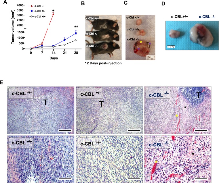
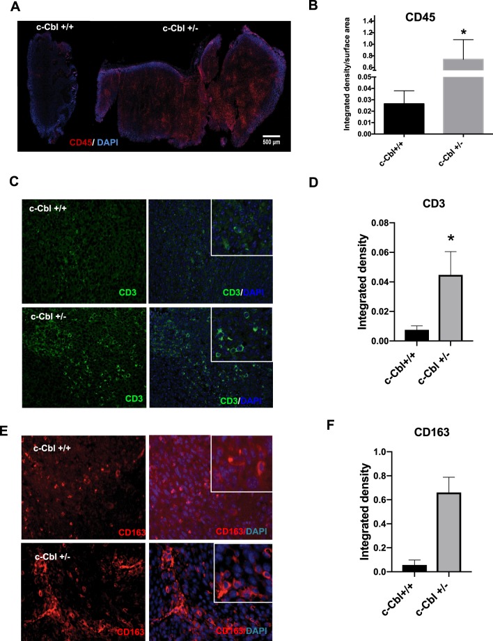
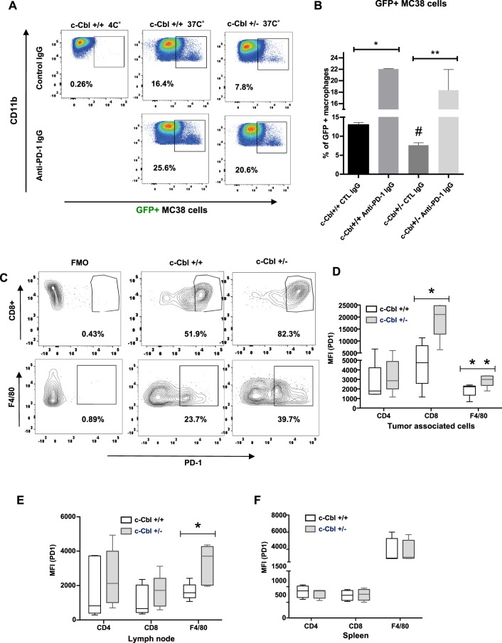
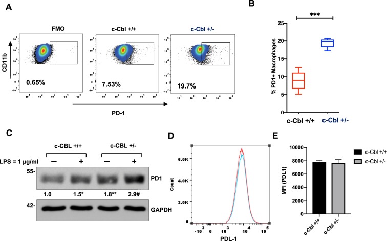
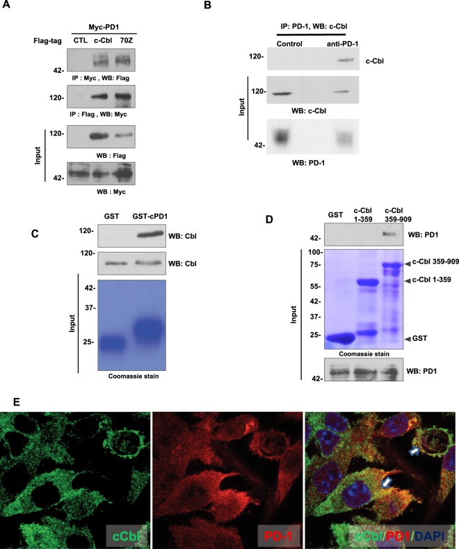
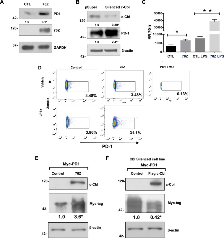
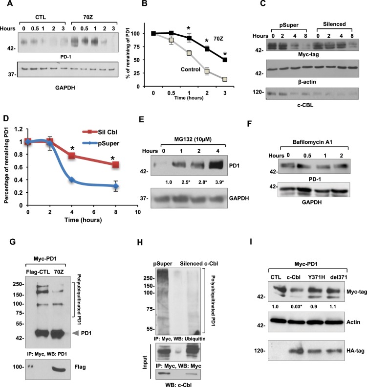
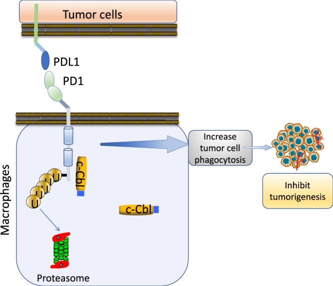
Casitas B lymphoma (c-Cbl) is an E3 ubiquitin ligase and a negative regulator of colorectal cancer (CRC). Despite its high expression in immune cells, the effect of c-Cbl on the tumor microenvironment remains poorly understood. Here we demonstrate that c-Cbl alters the tumor microenvironment and suppresses Programmed cell death-1 (PD-1) protein, an immune checkpoint receptor. Using syngeneic CRC xenografts, we observed significantly higher growth of xenografts and infiltrating immune cells in c-Cbl+/- compared to c-Cbl+/+ mice. Tumor-associated CD8+ T-lymphocytes and macrophages of c-Cbl+/- mice showed 2-3-fold higher levels of PD-1. Functionally, macrophages from c-Cbl+/- mice showed a 4-5-fold reduction in tumor phagocytosis, which was restored with an anti-PD-1 neutralizing antibody suggesting regulation of PD-1 by c-Cbl. Further mechanistic probing revealed that C-terminus of c-Cbl interacted with the cytoplasmic tail of PD-1. c-Cbl destabilized PD-1 through ubiquitination- proteasomal degradation depending on c-Cbl's RING finger function. This data demonstrates c-Cbl as an E3 ligase of PD-1 and a regulator of tumor microenvironment, both of which were unrecognized components of its tumor suppressive activity. Advancing immune checkpoint and c-Cbl biology, our study prompts for probing of PD-1 regulation by c-Cbl in conditions driven by immune checkpoint abnormalities such as cancers and autoimmune diseases.


Conflict of interest statement

The authors declare no competing interests.

Figures

Figure 1
Figure 1
Reduced c-Cbl activity enhances tumor growth and immune infiltrates. (A) Average of tumor volumes from six mice injected subcutaneously with MC38 cells in c-Cbl+/+ and c-Cbl+/− groups and five in the c-Cbl−/− group. All the c-Cbl−/− mice had to be euthanized by 10–12 days with rapid development of the xenograft along with ulceration on the tumor. Student’s t-test was performed. Error bars = SEM. *p < 0.0001 corresponds to growth of tumors in c-Cbl−/− mice compared to both c-Cbl+/− and c-Cbl+/+ groups. **p = 0.003 compares the group tumor growth c-between Cbl+/− and c-Cbl−/− group. (B) A representative mouse from three groups is shown at 12 days post-injection. (C) Excised xenografts are shown. Blue asterisk corresponds to the areas of necrosis and liquefaction (soft by palpation) compared to other areas of xenografts and yellow asterisk demarcates areas of ulceration. (D) Excised MC38 xenografts are shown. Representative images of a pair of c-Cbl+/+ and c-Cbl+/− mice are shown. (E) H&E stain of xenografts from different groups of mice are shown at 40X and 100X magnification. Xenografts from c-Cbl+/+ and c-Cbl+/− mice showed tumor cells (T) without evidence of acellular areas with fragmented nuclei suggestive of necrosis. Xenografts from c-Cbl−/− mice showed acellular areas suggestive of necrosis (marked with a black asterisk) and several areas of pools of RBCs suggestive of hemorrhage (marked with a yellow asterisk). Scale bar = 25 micron (40×) and 50 micron (100×).
Figure 2
Figure 2
Increased immune infiltrate in xenografts of c-Cbl+/− mice. (A) Xenografts were stained for CD45+ cells and DAPI for the nuclei. Intense DAPI stain at the periphery of the tumor is due to increase staining at the edges (edge effect). Representative Images from six xenografts per group. (B) CD45+ cell infiltration was quantitated as integrated density and normalized for the area of the tumor in square micron. Average of four sections of the tumor per mouse is shown. Total of six tumors per group. Student’s t-test was performed. Error bars = SEM. p = 0.043. (C) Xenograft from each mouse was stained for CD3+ cells. The insert shows tumor-associated lymphocytes. DAPI stained nuclei. Representative images from six xenografts per group are shown. (D) CD3+ cell infiltration was quantitated as integrated density. Average of ten sections of the tumor per mouse is shown. Total of six tumors per group. Student’s t-test was performed. Error bars = SEM. p = 0.03. (E) Xenograft from each mouse was stained for CD163+ cells. The insert shows the tumor-associated macrophages. Representative images from six xenografts per group are shown. (F) CD163+ cell infiltration was quantitated as integrated density. Average of ten sections of the tumor per mouse is shown. Total of six tumors per group. Student’s t-test was performed. Error bars = SEM. P < 0.001.
Figure 3
Figure 3
Reduced c-Cbl activity compromises tumor phagocytosis while increasing PD-1 expression in macrophages. (A) BMDM differentiated from c-Cbl+/+ and c-Cbl+/− for 7 days were co-cultured with GFP+ MC38 tumor cells for 4 hours and treated with iso-type control or anti-PD-1 neutralizing antibodies. Representative FACS from three independent experiments is shown. Gate set with GFP and Fluorescence Minus One (FMO) served as control. (B) Averages from three independent experiments with MC38 cells are shown. 2-way ANOVA and Student’s t-test were performed. Error bars = SEM. #p = 0.013 compares c-Cbl+/+ and c-Cbl+/− macrophages treated with the control antibody, *p = 0.002, **p = 0.05. (C) MC38 xenografts were harvested and the tumor-associated immune cells were sorted for live and dead cells using Zombie dye followed by cell-type specific markers such as CD8 for cytotoxic T-lymphocytes and F4/80 for activated macrophages. The Fluorescence Minus One Control or FMO served as control. Representative FACS plots from six independent tumors are shown. Gates set with PD-1 Fluorescence Minus One (FMO) control. (D) Mean fluorescence intensity (MFI) of PD-1 on tumor-associated immune cells from six mice in each group is shown as box plots. The borders of the box depict 25th and 75th percentile and whisker corresponding to minimum and maximum values. Student’s t-test were performed. *p = 0.007, **p = 0.050. (E) MFI of PD-1 on cells from lymph nodes of tumor-injected six mice in each group is shown. Student’s t-test were performed. * p = 0.002. (F) MFI of PD-1 from splenic cells from un-injected mice six mice per group. Error bars = SEM.
Figure 4
Figure 4
Bone marrow derived c-Cbl+/− macrophages express higher levels of PD-1. (A) BMDM differentiated from c-Cbl+/+ and c-Cbl+/− for 7 days were subjected to FACS analysis. The cells were gated using CD11b and PD-1. Percentage of PD-1+ macrophages are shown. Fluorescence minus one (FMO) served as a control. Representative FACS plots from three independent experiments are shown. (B) Average from three independent experiments is shown. Student’s t-test were performed. Error bars = SEM. *p < 0.001. (C) Lysates of BMDMs from mice differentiated for 7 days and treated with LPS overnight. Representative immunoblots from three independent experiments. Student’s t-test with Bonferroni correction was performed. *p = 0.004 for PD-1 in c-Cbl+/+ macrophages with and without LPS, **p = 0.001 for PD-1 between c-Cbl+/+ and c-Cbl+/− macrophages without LPS and #p = 0.02 for LPS-treated c-Cbl+/+ and c-Cbl+/− macrophages. Here, and in all other blots, molecular weights in kilo Dalton are denoted on the left. (D) BMDM from c-Cbl+/+ (red line) and c-Cbl+/− (blue line) mice differentiated for 7 days and treated with LPS overnight were examined using FACS for PDL-1. Representative MFI of PDL-1 from three independent pairs of mice is shown. (E) Average MFI of PDL-1 on BMDM obtained from both the groups (N = 3 mice/group) is shown. Student’s t-test were performed. Error bars = SEM. p = 0.3.
Figure 5
Figure 5
Cytoplasmic tail of PD-1 interacts with the C-terminus of c-Cbl to suppress PD-1. (A) Lysates of HEK293T cells co-expressing Myc-tagged PD-1 and Flag-tagged-c-Cbl or 70Z-c-Cbl were immunoprecipitated with Myc-tagged antibody and probed using Flag-tag antibody and vice versa. Five percent of inputs were probed separately. Representative images of three independent experiment are shown. (B) Lysates of RAW264.7 macrophages cells were immunoprecipitated with PD-1 antibody and probed using c-Cbl antibody. Five percent of inputs were probed separately. Representative images of two independent experiments are shown. (C) Recombinant GST-tagged intracytoplasmic tail of PD-1 was treated with lysates from RAW267.4. The eluents were probed for c-Cbl. Inputs were probed separately. The GST-beads were stained using Commassie dye. Representative immunoblots of three experiments are shown. (D) Recombinant GST-tagged c-Cbl 1-359 and 359–909 were treated with lysates from RAW264.7 cells. The eluents were probed for PD-1. 5% of lysates and the beads stained with Commassie are shown as inputs. Representative immunoblots of three experiments are shown. (E) Laser confocal microscopy was performed on RAW264.7 cells fixed and stained for c-Cbl and PD-1. Representative images from 100 randomly counted cells are shown. Scale bar = 10 μM.
Figure 6
Figure 6
c-Cbl suppresses PD-1. (A) RAW264.7 cells transfected with Myc-tagged 70Z-c-Cbl and control (CTL) plasmids were probed separately. Representative immunoblots of five experiments are shown. Student’s t-test was performed. *p = 0.003. (B) RAW267.4 cells expressing control (pSuper) or c-Cbl shRNA (silenced c-Cbl) were probed. Equal amounts of lysates were probed separately for PD-1. Representative immunoblots from three independent experiments. Student’s t-test was performed. * and **p = 0.001. (C) RAW267.4 cells transfected with 70Z-c-Cbl and control plasmids were treated for 16 hours with LPS (1 ug/ml) followed by FACS analysis. The live cells using Zombie were gated for PD-1. MFI of PD-1+ cells from three independent experiments is shown. ANOVA and Student’s t-test were performed. Error bars = SD. *p = 0.03 and **p = 0.001. (D) RAW267.4 expressing 70Z-c-Cbl and control plasmids were treated overnight with or without LPS (1 ug/ml) for 16 hours followed by FACS analysis. The cells were gated using Zombie dye and for PD-1. Gates set with PD-1 Fluorescence Minus One (FMO) control. Representative FACS plots from three independent experiments are shown. (E) HEK 293 T cells co-expressing Myc-tagged PD-1 and different c-Cbl constructs were lyzed and probed for c-Cbl and actin. Equal amounts of lysates were probed for Myc-tagged PD-1 given close proximity to Actin. A representative of three experiments is shown. Increase in PD-1 was compared using Student’s t-test. *p = 0.01. (F) HEK2913T cells silenced for c-Cbl were transfected with Myc-PD-1 and c-Cbl or control vector. The lysates were probed for c-Cbl and Actin. Equal amounts of lysates were probed for Myc-tagged PD-1 given close proximity to Actin. A representative of three experiments is shown. Suppression in PD-1 expression was compared using Student’s t-test. *p = 0.02.
Figure 7
Figure 7
c-Cbl destabilizes PD-1 and targets it for ubiquitination and proteasomal degradation. (A) RAW264.7 expressing control (CTL) and 70Z-c-Cbl were treated with Cycloheximide (300 μM) for the indicated time. Representative immunoblots from four experiments are shown. (B) Densitometry of PD-1 normalized to GAPDH and represented as the percentage of PD-1 at time = 0. Time to reach 50% of PD-1 was considered as its half-life. Average of four experiments is shown. Error bars = SEM. Student’s t-test was used. *p = 0.03 and **p = 0.01. (C) Lysates from HEK-293T cells stably expressing control (pSuper) and c-Cbl shRNA (Silenced) vectors were transfected with Myc-PD-1 and treated with Cycloheximide (100 μM) for the indicated time. The amount of remaining PD-1 was detected by Western blotting. Equal amounts of lysates were probed separately to confirm c-Cbl silencing. A representative figure from four independent experiments is shown. (D) Densitometry of normalized PD-1 bands performed using ImageJ represented as the percentage of PD-1 at time 0 is shown. The time to reach 50% of initial PD-1 was considered as the half-life of PD-1. An average of four experiments is shown. Error bars = SD. Student’s t-test was performed. *p = 0.001 and **p = 0.003. (E) RAW264.7 cells treated with MG132 for indicated amount of time. Representative immunoblots from four independent experiments are shown. Student’s t-test with Bonferroni correction was used. Compared to time = 0, *p = 0.04 at 1 hour, p = 0.001 at 2 and 4 hours. (F) Raw264.7 cells treated with 200 nM of Baflomycin A1 for the indicated time points. The lysates were probed as shown. A representative of two experiments is shown. (G) HEK 293T cells expressing control (Flag-CTL) and 70Z-c-Cbl were pre-treated with 10 µM MG132 overnight and immunoprecipitated using PD-1 antibody. The eluents were probed with PD-1. 5% of lysates were probed separately with Flag-tag. Representative immunoblots from three experiments are shown. (H) HEK293T cells stably expressing shRNA of c-Cbl (Silenced c-Cbl) and control (pSuper) and transfected with Myc-tagged PD-1 were immunoprecipitated using Myc antibody. Eluents were probed with anti-ubiquitin antibody. 5% of lysates were probed for c-Cbl. Representative immunoblots from three experiments are shown. (I) HEK293T co-expressing Myc-tagged PD-1 and HA-tagged c-Cbl or control (CTL) constructs were probed and normalized to actin. Equal amounts of lysates were probed for HA-tag. Representative immunoblots from three independent experiments. 2-way ANOVA with multiple comparison and Student’s t-test were performed. Compared to CTL, *p = 0.02 for c-Cbl.
Figure 8
Figure 8
Model of c-Cbl mediated PD-1 regulation. c-Cbl in immune cells (such as macrophages) binds and ubiquitinates PD-1 to target it for proteasomal degradation. This results in downregulation of surface expression of PD-1, which in macrophages augments the tumor cell phagocytosis resulting in suppression of tumor growth.

References

    1. Siegel, R. L. et al. Colorectal Cancer Incidence Patterns in the United States, 1974–2013. J Natl Cancer Inst109, 10.1093/jnci/djw322 (2017). - PMC - PubMed
    1. Fearon, E. R. & Vogelstein, B. A genetic model for colorectal tumorigenesis. Cell 61, 759–767, 0092-8674(90)90186-I (1990). - PubMed
    1. Thien CB, Langdon WY. Cbl: many adaptations to regulate protein tyrosine kinases. Nat Rev Mol Cell Biol. 2001;2:294–307. doi: 10.1038/3506710035067100. - DOI - PubMed
    1. Schmidt, M. H. & Dikic, I. The Cbl interactome and its functions. Nat Rev Mol Cell Biol6, 907–918, nrm1762 10.1038/nrm1762 (2005). - PubMed
    1. Naramura M, Kole HK, Hu RJ, Gu H. Altered thymic positive selection and intracellular signals in Cbl-deficient mice. Proc Natl Acad Sci USA. 1998;95:15547–15552. doi: 10.1073/pnas.95.26.15547. - DOI - PMC - PubMed

Publication types

MeSH terms

Substances