. 2019 Dec 27;9(1):20002.
doi: 10.1038/s41598-019-56628-z.

An acute session of motor imagery training induces use-dependent plasticity

Affiliations

An acute session of motor imagery training induces use-dependent plasticity

Célia Ruffino et al. Sci Rep. .

Abstract

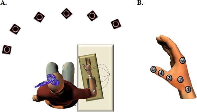
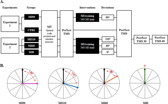
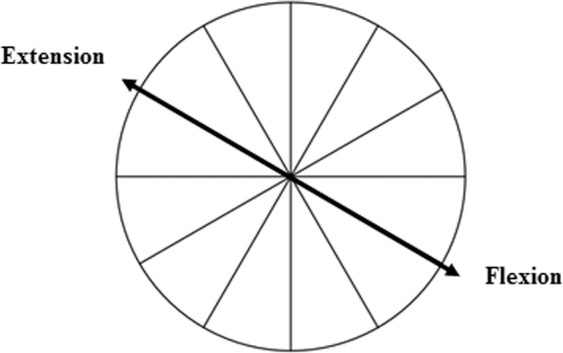
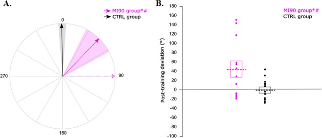
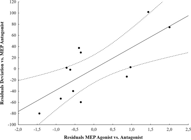
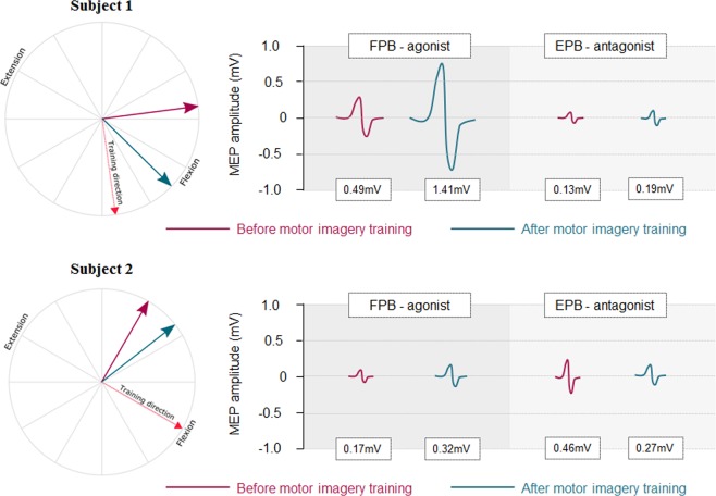
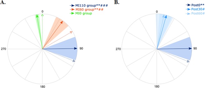
Motor imagery, defined as the mental representation of an action without movement-related sensory inputs, is a well-known intervention to improve motor performance. In the current study, we tested whether use-dependent plasticity, a mechanism underlying motor learning, could be induced by an acute session of motor imagery. By means of transcranial magnetic stimulation (TMS) over the left primary motor cortex, we evoked isolated thumb movements in the right hand and assessed corticospinal excitability in the flexor and extensor pollicis brevis muscles. We measured the mean TMS-induced movement direction before and after an acute session of motor imagery practice. In a first experiment, participants of the imagery group were instructed to repeatedly imagine their thumb moving in a direction deviated by 90° from the pre-test movement. This group, but not the control group, deviated the post-training TMS-induced movements toward the training target direction (+44° ± 62° and -1° ± 23°, respectively). Interestingly, the deviation magnitude was driven by the corticospinal excitability increase in the agonist muscle. In a second experiment, we found that post-training TMS-induced movements were proportionally deviated toward the trained direction and returned to baseline 30 minutes after the motor imagery training. These findings suggest that motor imagery induces use-dependent plasticity and, this neural process is accompanied by corticospinal excitability increase in the agonist muscle.


Conflict of interest statement

The authors declare no competing interests.

Figures

Figure 1
Figure 1
(A) Position of the participant and the infra-red cameras. (B) Position of retro-reflective markers.
Figure 2
Figure 2
(A) Intervention design. (B) Illustration of the target angles used to train each group. The black arrow pictures the mean TMS-induced movement at PreTest and the colored arrow represents the direction of the imagined movements during training. Note that mean direction of PreTest TMS-induced movements varied across participants. MI = Motor Imagery; MT = Movement Threshold; TMS = Transcranial Magnetic Stimulation.
Figure 3
Figure 3
Illustration of actual brisk flexion and extension. When the arm and forearm were positioned in the cast, thumb flexion and extension were oriented about 60° counterclockwise from the vertical axis. This helped determining for each individual the role (agonist or antagonist) of flexor and extensor pollicis brevis muscles relative to the trained direction.
Figure 4
Figure 4
Post-training deviation. (A) For each group, the solid arrow and the colored area correspond to the mean normalized deviation and to the standard error around the mean, respectively. The dotted pink arrow reminds the training direction for the motor imagery group. All participants are represented in a similar frame of reference, in which 0 is the TMS-induced movement direction at PreTest. (B) For each group, the dotted lines correspond to the mean normalized deviation. The boxes represent the standard error of the mean and the dots refer to individual values. *Significantly different from PreTest (P < 0.05); #significant difference between MI90 and CTRL group (P < 0.05).
Figure 5
Figure 5
Partial scatter plot of the association between the movement deviation and the ratio MEP Agonist when controlling for the ratio MEP Antagonist (partial correlation = 0.74, P = 0.009). Solid line = fitting regression line; dotted line = 95% confidence regression bands.
Figure 6
Figure 6
Typical representation of TMS-induced movement direction and MEP amplitude in flexor pollicis brevis (FPB) and extensor pollicis brevis (EPB) muscles in two subjects of the MI90 group. In both subjects, FPB was the agonist of the trained movement. Subject 1 deviated from Pre to PostTest by +52° and increased MEP amplitude in the agonist and antagonist muscles by 215% and 51%, respectively. Subject 2 deviated from Pre to PostTest by +22° and increased and decreased MEP amplitude in the agonist and antagonist muscles by 88% and 42%, respectively.
Figure 7
Figure 7
(A) Post-training deviation. For each group, the solid arrow and the colored area correspond to the mean normalized deviation and to the standard error around the mean, respectively. The dotted arrow reminds the training direction for each group. All participants are represented in a similar frame of reference, in which 0 is the TMS-induced movement direction at PreTest. **Significantly different from PreTest for MI60 and MI110 (P = 0.01 and P = 0.003 respectively); ##significant different between MI60 and MI0 (P = 0.01) and ###significant difference between MI110 and MI0 (P < 0.001). (B) Lasting effects of use-dependent plasticity for the MI110 group. *And **significantly different from PreTest (P < 0.05 and P < 0.01, respectively); #and ###significantly different from MI110 Post-test0 (P < 0.05 and P < 0.001, respectively).

References

    1. Diedrichsen J, White O, Newman D, Lally N. Behavioral/Systems/Cognitive Use-Dependent and Error-Based Learning of Motor Behaviors. J. Neurosci. 2010;30:5159–5166. doi: 10.1523/JNEUROSCI.5406-09.2010. - DOI - PMC - PubMed
    1. Verstynen T, Sabes PN. How each movement changes the next: an experimental and theoretical study of fast adaptive priors in reaching. J. Neurosci. 2011;31:10050–9. doi: 10.1523/JNEUROSCI.6525-10.2011. - DOI - PMC - PubMed
    1. Classen J, Liepert J, Wise SP, Hallett M, Cohen LG. Rapid plasticity of human cortical movement representation induced by practice. J. Neurophysiol. 1998;79:1117–23. doi: 10.1152/jn.1998.79.2.1117. - DOI - PubMed
    1. Bütefisch C, Davis B, Wise S. Mechanisms of use-dependent plasticity in the human motor cortex. PNAS. 2000;97:3661–5. doi: 10.1073/pnas.97.7.3661. - DOI - PMC - PubMed
    1. Celnik P, et al. Encoding a motor memory in the older adult by action observation. NeuroImage. 2006;29:677–84. doi: 10.1016/j.neuroimage.2005.07.039. - DOI - PubMed

Publication types