. 2019 Dec 27;9(1):19940.
doi: 10.1038/s41598-019-56507-7.

Computational assessment of the retinal vascular tortuosity integrating domain-related information

Affiliations

Computational assessment of the retinal vascular tortuosity integrating domain-related information

L Ramos et al. Sci Rep. .

Abstract

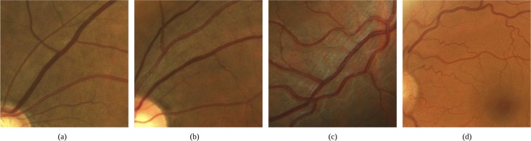
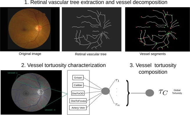
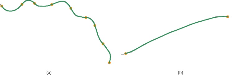
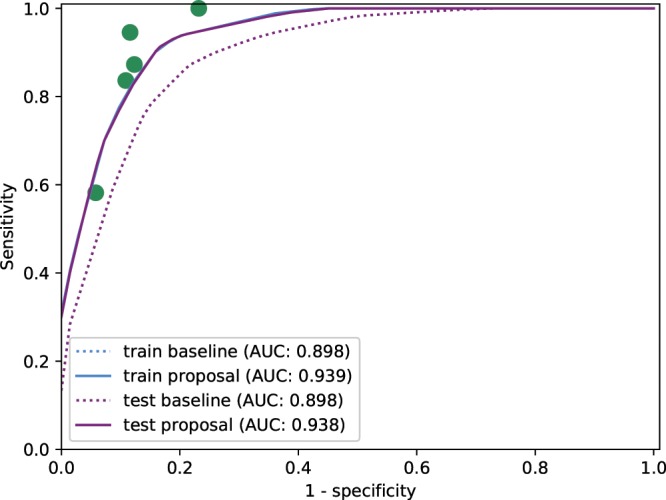
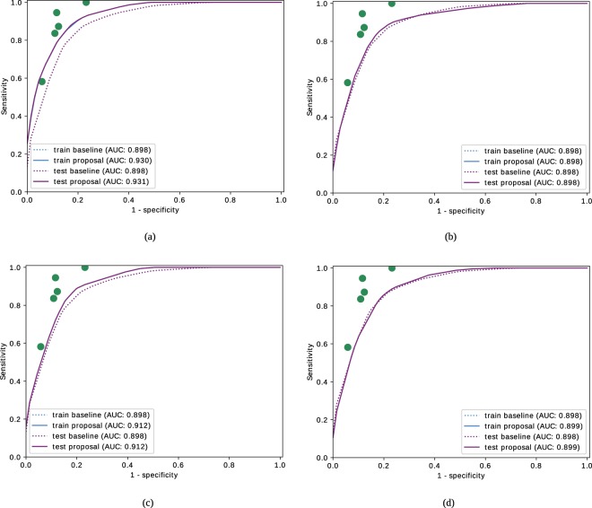
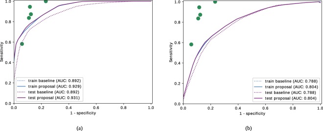
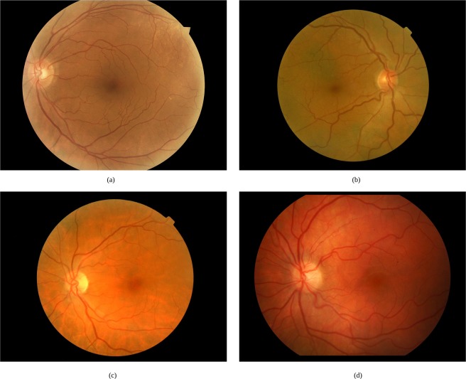
The retinal vascular tortuosity presents a valuable potential as a clinical biomarker of many relevant vascular and systemic diseases. Commonly, the existent approaches face the tortuosity quantification by means of fully mathematical representations of the vessel segments. However, the specialists, based on their diagnostic experience, commonly analyze additional domain-related information that is not represented in these mathematical metrics of reference. In this work, we propose a novel computational tortuosity metric that outperforms the mathematical metrics of reference also incorporating anatomical properties of the fundus image such as the distinction between arteries and veins, the distance to the optic disc, the distance to the fovea, and the vessel caliber. The evaluation of its prognostic performance shows that the integration of the anatomical factors provides an accurate tortuosity assessment that is more adjusted to the specialists' perception.


Conflict of interest statement

The authors declare no competing interests.

Figures

Figure 1
Figure 1
Retinal images with (a,b) non-tortuous and (c,d) tortuous blood vessels.
Figure 2
Figure 2
Steps for the computational tortuosity measurement. The first block is in charge of the extraction of the arterio-venous tree and the decomposition into its constituent vessels. In the second block, a local tortuous value is computed for each vessel. In the third block, the local tortuosity values are combined to compute the global tortuosity quantification.
Figure 3
Figure 3
Grisan Vessel decomposition into the constituent subsegments of constant-sign curvature.
Figure 4
Figure 4
ROC curves for the baseline metric τG and the proposed metric τC that includes the anatomical factors in the training and test processes.
Figure 5
Figure 5
ROC curves for the baseline metric and proposed metric using independently each individual factor in the training and test process. The considered anatomical factors are (a) the distinction between arteries and veins, (b) the distance to the optic disc, (c) the distance to the fovea and (d) the vessel caliber.
Figure 6
Figure 6
ROC curves for the baseline metric and proposed metric considering separately (a) arteries and (b) veins in the training and test process.
Figure 7
Figure 7
Examples of representative cases where the incorporation of the anatomical factors allows to produce a correct tortuosity characterization.

Similar articles

Cited by

References

    1. Witt Nicholas, Wong Tien Y., Hughes Alun D., Chaturvedi Nish, Klein Barbara E., Evans Richard, McNamara Mary, Thom Simon A. McG, Klein Ronald. Abnormalities of Retinal Microvascular Structure and Risk of Mortality From Ischemic Heart Disease and Stroke. Hypertension. 2006;47(5):975–981. doi: 10.1161/01.HYP.0000216717.72048.6c. - DOI - PubMed
    1. Dougherty G, Johnson MJ, Wiers MD. Measurement of retinal vascular tortuosity and its application to retinal pathologies. Medical & Biological Engineering & Computing. 2009;48:87. doi: 10.1007/s11517-009-0559-4. - DOI - PubMed
    1. lui Cheung CY, et al. Retinal vascular tortuosity, blood pressure, and cardiovascular risk factors. Ophthalmology. 2011;118:812–818. doi: 10.1016/j.ophtha.2010.08.045. - DOI - PubMed
    1. Sasongko MB, et al. Retinal arteriolar tortuosity is associated with retinopathy and early kidney dysfunction in type 1 diabetes. American Journal of Ophthalmology. 2012;153:176–183.e1. doi: 10.1016/j.ajo.2011.06.005. - DOI - PubMed
    1. Kalitzeos AA, Lip GYH, Heitmar R. Retinal vessel tortuosity measures and their applications. Experimental eye research. 2013;106:40–6. doi: 10.1016/j.exer.2012.10.015. - DOI - PubMed

Publication types