Review
. 2019 Nov;292(1):149-163.
doi: 10.1111/imr.12823.

T-cell exhaustion in HIV infection

Affiliations
Review

T-cell exhaustion in HIV infection

Craig Fenwick et al. Immunol Rev. 2019 Nov.

Abstract

The T-cell response is central in the adaptive immune-mediated elimination of pathogen-infected and/or cancer cells. This activated T-cell response can inflict an overwhelming degree of damage to the targeted cells, which in most instances leads to the control and elimination of foreign invaders. However, in conditions of chronic infection, persistent exposure of T cells to high levels of antigen results in a severe T-cell dysfunctional state called exhaustion. T-cell exhaustion leads to a suboptimal immune-mediated control of multiple viral infections including the human immunodeficiency virus (HIV). In this review, we will discuss the role of T-cell exhaustion in HIV disease progression, the long-term defect of T-cell function even in aviremic patients on antiretroviral therapy (ART), the role of exhaustion-specific markers in maintaining a reservoir of latently infected cells, and exploiting these markers in HIV cure strategies.

Keywords: HIV; PD-1; T-cell exhaustion; chronic viral infection; immune checkpoint inhibitors.


Conflict of interest statement

All authors declare no conflict of interest with this review.

Figures

Figure 1
Figure 1
T‐cell immune checkpoint inhibitors and related ligands. TCR interaction with the antigenic peptide‐MHC complex displayed by professional APCs delivers the primary signal for T‐cell activation. The CD28 co‐receptor and other costimulatory receptors enhance the T‐cell stimulatory signal following interaction with their corresponding ligands. Immune checkpoint inhibitors including PD‐1, CTLA‐4, LAG3, TIGIT, TIM3, CD160, and 2B4 act to suppress T‐cell signal following interaction with their related ligands expressed on APCs
Figure 2
Figure 2
HIV Infection and progression of exhaustion. During the acute phase of HIV infection, the majority of individuals experience a dramatic increase in HIV viral load and increased levels of the PD‐1 receptor on HIV‐specific T cells. In contrast, elite controllers maintain low viral load levels and consequently, T cells express low levels of PD‐1. Untreated patients with high viral load progress to the chronic phase of HIV infection where persistent elevated levels of HIV antigen result in T‐cell exhaustion with high expression levels of PD‐1. ART in most patients significantly inhibits viral replication, resulting in decreased plasma viral load to levels below the limit of detection with standard assays. With low levels of viral antigen present, PD‐1 expression decreases to lower levels on HIV‐specific T cells
Figure 3
Figure 3
Phenotype, transcriptional, and functional profile of T cells progressing to exhaustion. Antigen‐specific stimulation of resting T cells leads to a functionally active T‐cell response with increased cell surface PD‐1, upregulated expression of the T‐bet transcription factor and a strong functional and proliferative response. Chronic stimulation with high levels of antigen drive T cells into an exhausted state characterized by high levels of PD‐1, an increased expression of additional immune checkpoint inhibitors and a pronounced T‐cell dysfunction. Compared to functionally active T cells, highly exhausted T cells have elevated expression of transcription factors including NFAT, Batf, Eomes, and Blimp‐1 with decreased levels of T‐bet
Figure 4
Figure 4
Signaling in T‐cell activation and PD‐1–mediated suppression. T‐cell activation through the TCR/CD3 complex leads to the phosphorylation of proximal signaling molecules that trigger downstream activation of NFAT, CREB, and p38 pathways. In the absence of a costimulatory signaling through CD28, TCR activation of T cells leads to a hyporesponsive, anergic state. Signaling through the CD28 costimulatory receptor enhances activation of the PI3K/AKT/NFκB pathways that support T‐cell proliferation and reduce apoptosis. Interaction of PD‐1 with PDL‐1 during T‐cell activation recruits the PD‐1/SHP2 tyrosine phosphatase complex into the vicinity of TCR/CD3 and the CD28 co‐receptor. This recruitment results in the dephosphorylation of membrane proximal PI3K, ZAP‐70 and the intracellular domains of CD28 and the CD3ζ chain, which suppresses T‐cell activation. In PD‐1 high exhausted T cells from chronically infected HIV donors, the ligation of PD‐1 with PDL‐1 results in reduced phosphorylation of TCR proximal Lck and Zap‐70 and downstream Erk1/2 in the Ras‐MEK1/2‐Erk1/2 pathway. PD‐1 suppression of T‐cell activation also results in reduced calcium mobilization that is upstream of CREB and NFAT transcription factors. In the CD28 costimulatory receptor pathway, ligation of PD‐1 with PDL‐1 significantly inhibits the PI3K/AKT/NFκB pathway with reduced phosphorylation of PDK1 and AKT
Figure 5
Figure 5
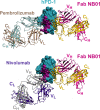
Antagonistic antibodies binding the PD‐1 receptor. The structure of human PD‐1 (hPD‐1) in complex with the anti‐PD‐1 NB01 Fab was solved by molecular replacement using crystals that diffracted to 2.2 Å resolution (6HIG). Molecular modeling by Cα superpositioning of hPD‐1 coordinates with the pembrolizumab (PDB 5GGS) and the nivolumab (PDB 5GGR) confirms that NB01 Fab binding to PD‐1 does not interfere with the binding of either pembrolizumab or nivolumab anti‐PD‐1 Abs. The hPDL‐1–binding surface (PDB 4ZQK) on hPD‐1 is colored in purple and is distinct from the binding epitope of the NB01 Fab that is non‐blocking of the PD‐1/PDL‐1 interaction
Figure 6
Figure 6
Anti‐PD‐1 antibody‐mediated restoration of exhausted T cells. (A) In the standard activation model of T cells expressing low levels of PD‐1, the immunological synapse forms with the TCR/CD3 complex at the core, surrounded by the CD28 costimulatory receptor within the cSMAC. This distribution forms a close complex of signaling molecules that enhances the T‐cell activation cascade. (B) In PD‐1 high T cells, PDL‐1 binding to PD‐1 recruits the PD‐1/SHP2 complex into the cSMAC that effectively suppresses signaling through the SHP2 phosphatase‐mediated dephosphorylation of TCR/CD3 and CD28 proximal signaling molecules. (C) Use of blocking anti‐PD‐1 antibodies such as pembrolizumab partially restores T‐cell signaling through limiting PD‐1/PDL‐1–mediated recruitment of the PD‐1/SHP2 complex into the cSMAC. The exclusion of SHP2 from the cSMAC reduces dephosphorylation of signaling molecules including ZAP‐70, Lck, PI3K, and AKT. However, our studies show that in activated T cells, pembrolizumab binding to PD‐1 pulls down a complex that includes SHP2, CD28, and PI3K following T‐cell activation. As such, PD‐1 may partially suppress T‐cell activation by recruiting CD28 away from the cSMAC and suppressing CD28 costimulation through the SHP2 phosphatase. (D) Our newly discovered anti‐PD‐1 antibody NB01 is non‐blocking of the PD‐1/PDL‐1 interaction and has equivalent antagonistic activity compared to pembrolizumab in restoring T‐cell signaling and antigen‐specific functional and proliferative activity to exhausted HIV‐specific T cells. Based on immunoprecipitation studies, our proposal is that non‐blocking anti‐PD‐1 antibodies act through inhibiting close contact of the PD‐1/SHP2 complex with CD28 and associated signaling molecules following T‐cell activation. As such, CD28 is free to migrate into the cSMAC and enhance T‐cell activation
Figure 7
Figure 7
Targeted killing of PD‐1–positive HIV‐infected cells. PD‐1 expressing CD4 T cells represent the primary compartment of HIV‐infected cells that produce replication competent virus. Targeted killing of these PD‐1 positive cells represents a novel therapeutic strategy that can deplete the HIV reservoir of infected cells. Antibody drug conjugates (ADC) use a toxin‐conjugated antibody that binds to the cell surface PD‐1 receptor. Internalization of PD‐1 results in ADC degradation within the lysosome, resulting in toxin release that specifically kills the PD‐1 expressing cell. An alternate strategy for the targeted killing of HIV infected PD‐1 positive cells is through antibody‐dependent cellular cytotoxicity (ADCC) with an IgG1 anti‐PD‐1 antibody. The Fc portion of the antibody binds to the FcγRIII receptors expressed on effector cells including natural killer (NK) cells. NK cells release cytotoxic granules that kill the PD‐1–positive HIV‐infected CD4 T cell

Similar articles

Cited by

References

    1. Schwartz RH. T cell anergy. Annu Rev Immunol. 2003;21:305‐334. - PubMed
    1. Chen L, Flies DB. Molecular mechanisms of T cell co‐stimulation and co‐inhibition. Nat Rev Immunol. 2013;13(4):227‐242. 10.1038/nri3405. - DOI - PMC - PubMed
    1. Wherry EJ, Kurachi M. Molecular and cellular insights into T cell exhaustion. Nat Rev Immunol. 2015;15(8):486‐499. 10.1038/nri3862. - DOI - PMC - PubMed
    1. Attanasio J, Wherry EJ. Costimulatory and Coinhibitory Receptor Pathways in Infectious Disease. Immunity. 2016;44(5):1052‐1068. 10.1016/j.immuni.2016.04.022. - DOI - PMC - PubMed
    1. Keir ME, Liang SC, Guleria I, et al. Tissue expression of PD‐L1 mediates peripheral T cell tolerance. J Exp Med. 2006;203(4):883‐895. 10.1084/jem.20051776. - DOI - PMC - PubMed

Publication types

MeSH terms