

Transcriptomic Analyses of Inner Ear Sensory Epithelia in Zebrafish
- PMID: 31883312
- PMCID: PMC8108486
- DOI: 10.1002/ar.24331
Transcriptomic Analyses of Inner Ear Sensory Epithelia in Zebrafish
Abstract
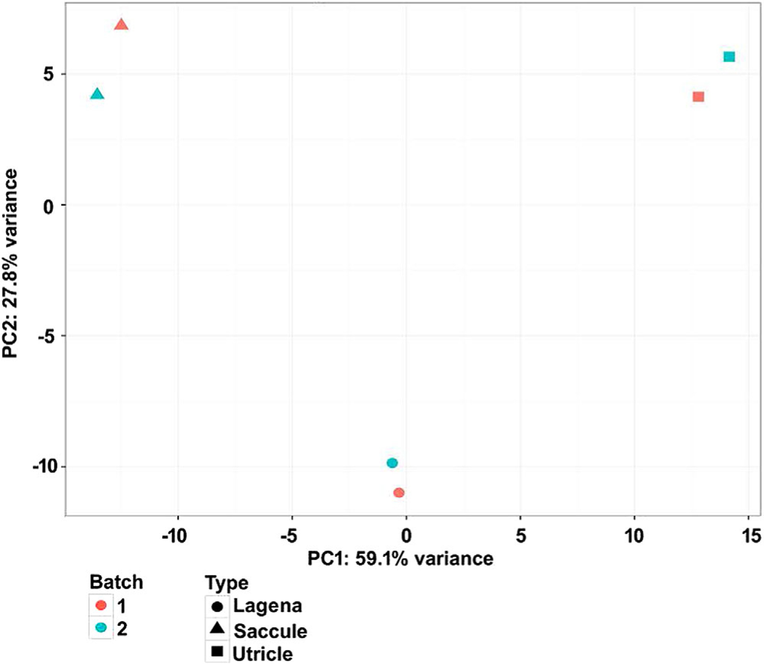
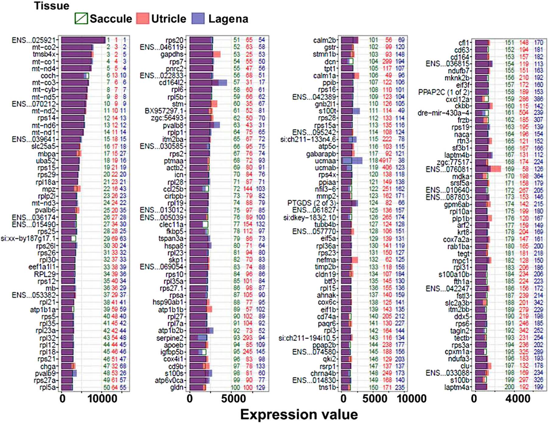
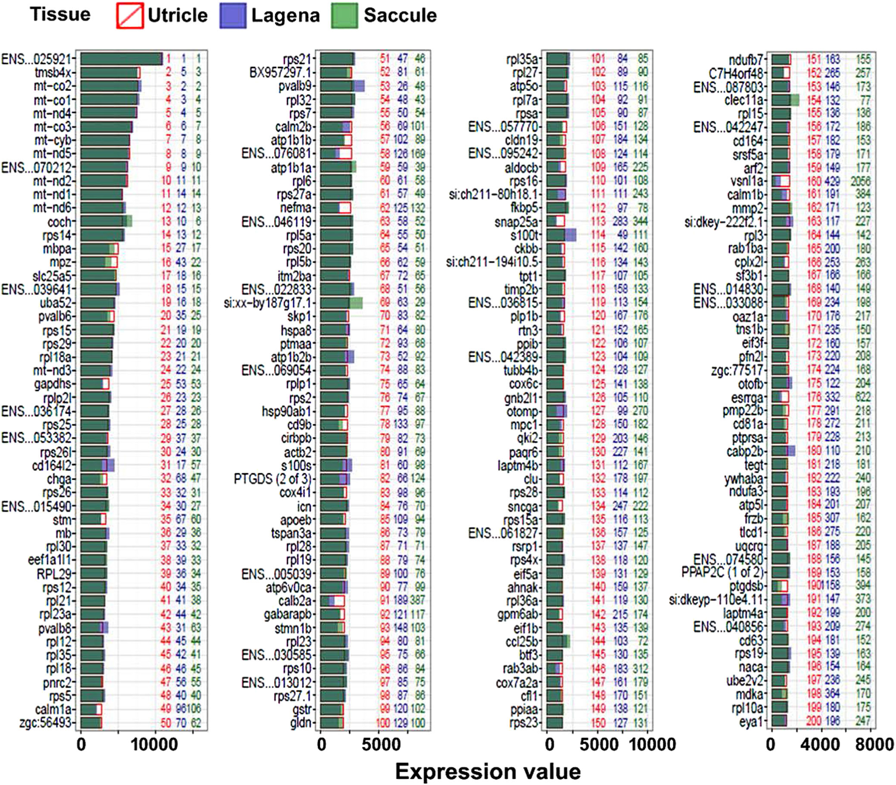
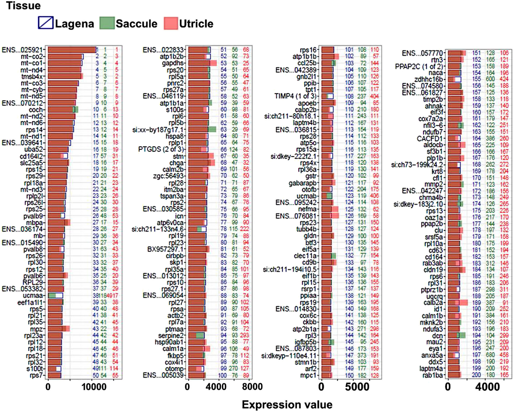
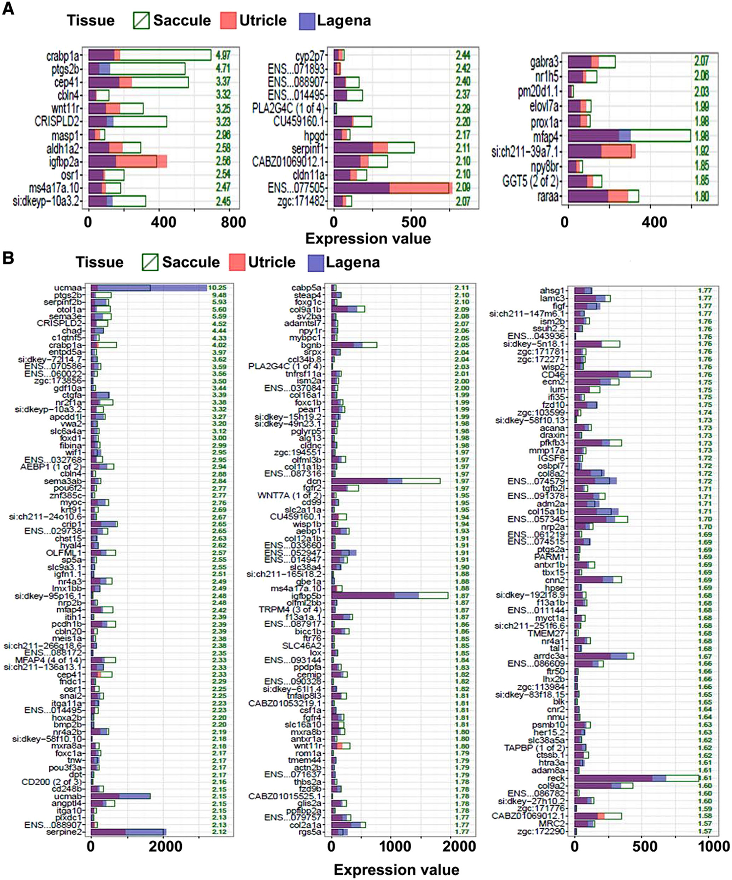
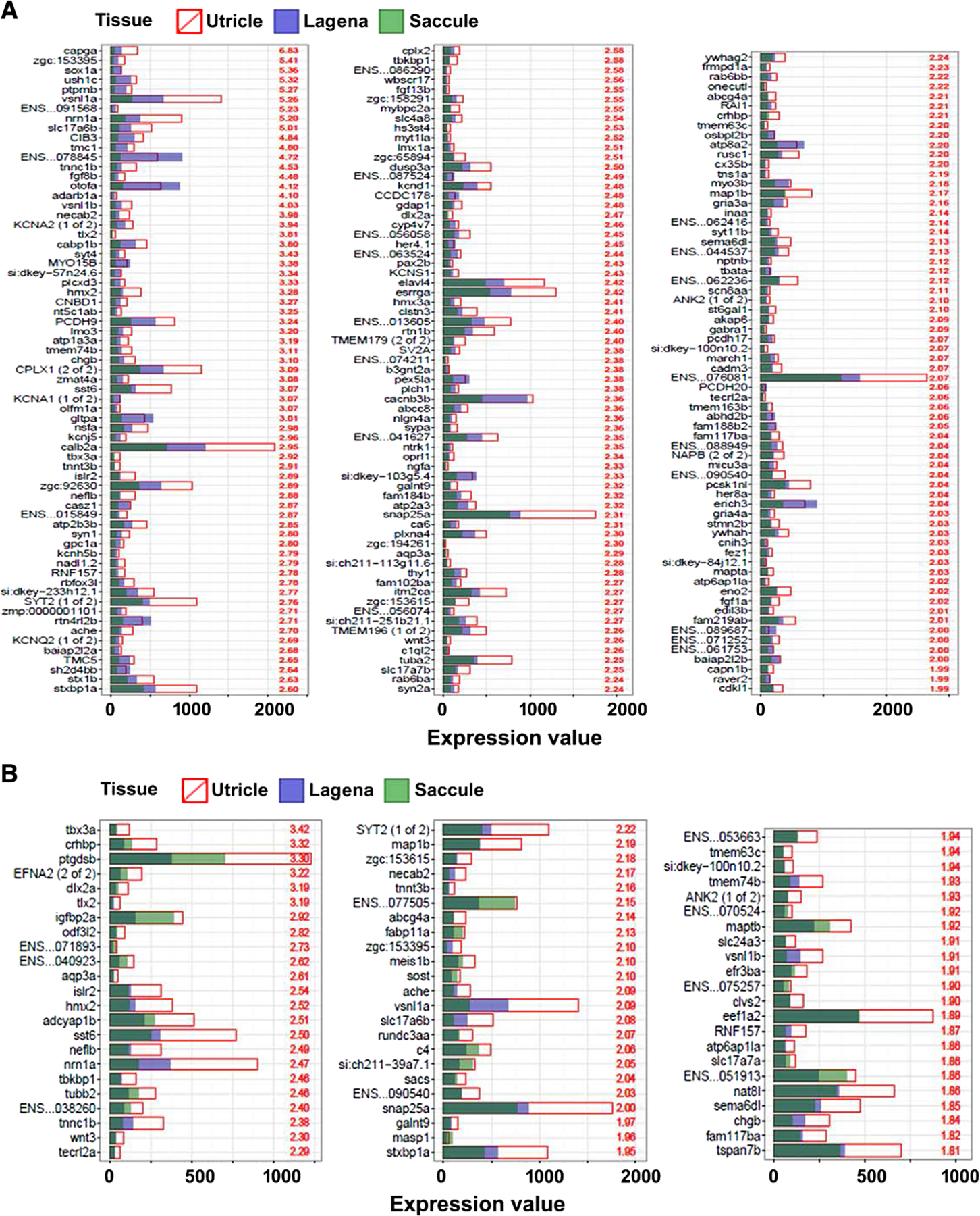
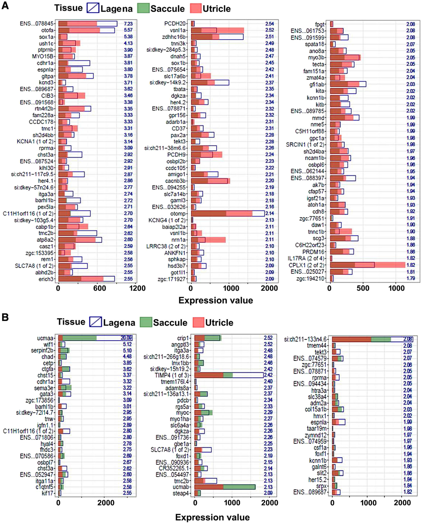
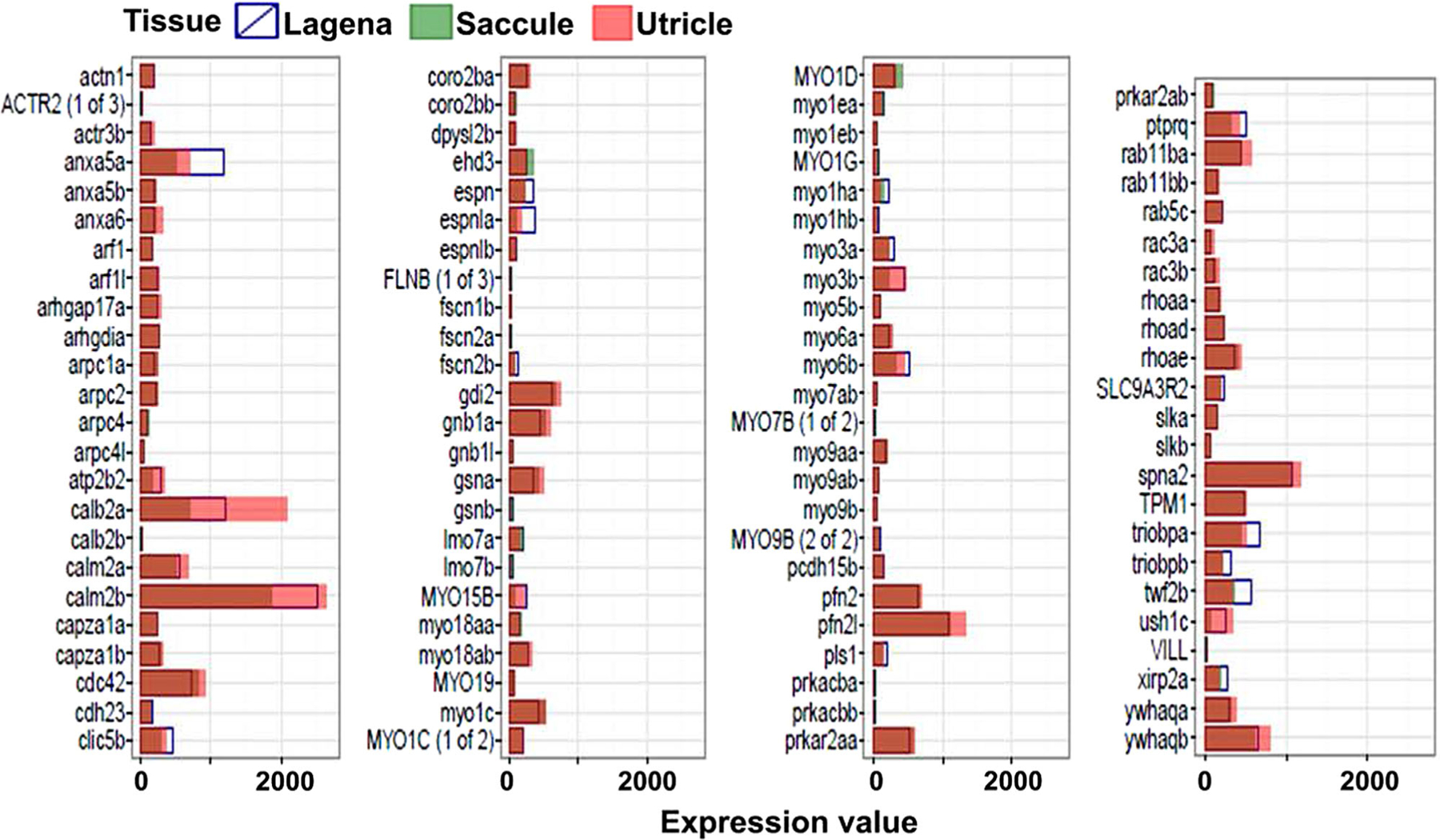
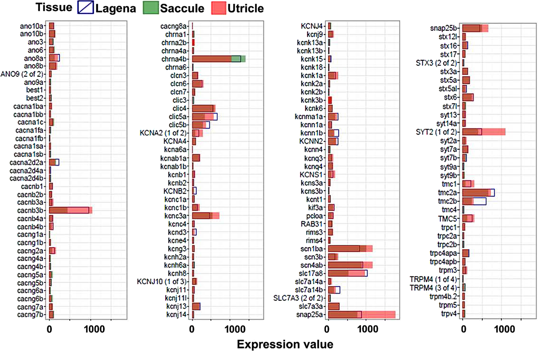
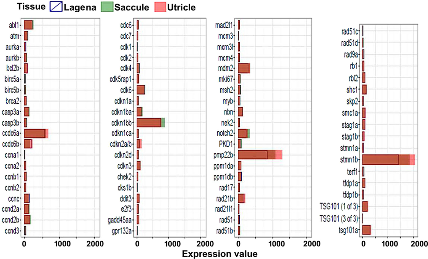
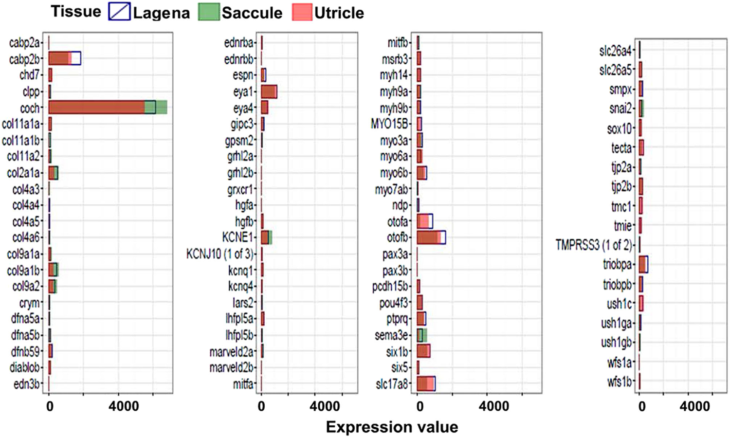
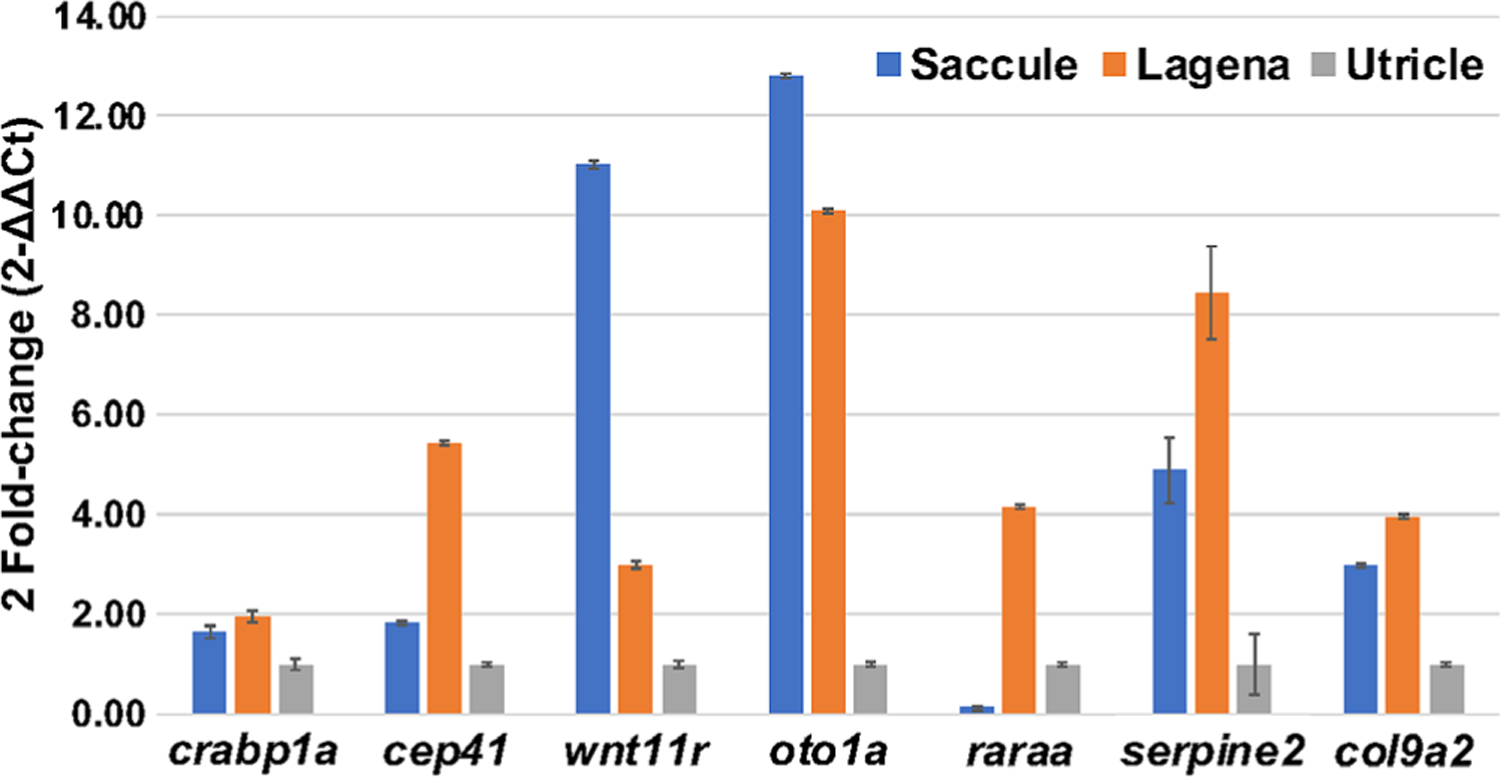
Analysis of gene expression has the potential to assist in the understanding of multiple cellular processes including proliferation, cell-fate specification, senesence, and activity in both healthy and disease states. Zebrafish model has been increasingly used to understand the process of hearing and the development of the vertebrate auditory system. Within the zebrafish inner ear, there are three otolith organs, each containing a sensory macula of hair cells. The saccular macula is primarily involved in hearing, the utricular macula is primarily involved in balance and the function of the lagenar macula is not completely understood. The goal of this study is to understand the transcriptional differences in the sensory macula associated with different otolith organs with the intention of understanding the genetic mechanisms responsible for the distinct role each organ plays in sensory perception. The sensory maculae of the saccule, utricle, and lagena were dissected out of adult Et(krt4:GFP)sqet4 zebrafish expressing green fluorescent protein in hair cells for transcriptional analysis. The total RNAs of the maculae were isolated and analyzed by RNA GeneChip microarray. Several of the differentially expressed genes are known to be involved in deafness, otolith development and balance. Gene expression among these otolith organs was very well conserved with less than 10% of genes showing differential expression. Data from this study will help to elucidate which genes are involved in hearing and balance. Furthermore, the findings of this study will assist in the development of the zebrafish model for human hearing and balance disorders. Anat Rec, 303:527-543, 2020. © 2019 American Association for Anatomy.
Keywords: hearing; microarray; neurosensory epithelium; transcriptome analysis; zebrafish inner ear.
© 2019 American Association for Anatomy.
Figures
Similar articles
-
The role of ear stone size in hair cell acoustic sensory transduction.Sci Rep. 2013;3:2114. doi: 10.1038/srep02114. Sci Rep. 2013. PMID: 23817603 Free PMC article.
-
The alcohol-sensitive period during early octavolateral organ development in zebrafish (Danio rerio).J Neurosci Res. 2017 May;95(5):1194-1203. doi: 10.1002/jnr.24017. Epub 2017 Jan 20. J Neurosci Res. 2017. PMID: 28105691
-
Hearing Assessment in Zebrafish During the First Week Postfertilization.Zebrafish. 2016 Apr;13(2):79-86. doi: 10.1089/zeb.2015.1166. Epub 2016 Jan 26. Zebrafish. 2016. PMID: 26982161 Free PMC article.
-
The genetics of hair-cell function in zebrafish.J Neurogenet. 2017 Sep;31(3):102-112. doi: 10.1080/01677063.2017.1342246. Epub 2017 Jul 13. J Neurogenet. 2017. PMID: 28705044 Free PMC article. Review.
-
Diversity of Inner Ears in Fishes: Possible Contribution Towards Hearing Improvements and Evolutionary Considerations.Adv Exp Med Biol. 2016;877:341-91. doi: 10.1007/978-3-319-21059-9_16. Adv Exp Med Biol. 2016. PMID: 26515322 Review.
Cited by
-
Small fish, big prospects: using zebrafish to unravel the mechanisms of hereditary hearing loss.Hear Res. 2020 Nov;397:107906. doi: 10.1016/j.heares.2020.107906. Epub 2020 Feb 6. Hear Res. 2020. PMID: 32063424 Free PMC article.
-
Optimizing gRNA selection for high-penetrance F0 CRISPR screening for interrogating disease gene function.Nucleic Acids Res. 2025 Feb 27;53(5):gkaf180. doi: 10.1093/nar/gkaf180. Nucleic Acids Res. 2025. PMID: 40103232 Free PMC article.
-
Single-Cell Sequencing Applications in the Inner Ear.Front Cell Dev Biol. 2021 Feb 12;9:637779. doi: 10.3389/fcell.2021.637779. eCollection 2021. Front Cell Dev Biol. 2021. PMID: 33644075 Free PMC article. Review.
-
The Genomics of Auditory Function and Disease.Annu Rev Genomics Hum Genet. 2022 Aug 31;23:275-299. doi: 10.1146/annurev-genom-121321-094136. Epub 2022 Jun 6. Annu Rev Genomics Hum Genet. 2022. PMID: 35667089 Free PMC article. Review.
-
A regulatory network of Sox and Six transcription factors initiate a cell fate transformation during hearing regeneration in adult zebrafish.Cell Genom. 2022 Sep 14;2(9):100170. doi: 10.1016/j.xgen.2022.100170. Epub 2022 Aug 22. Cell Genom. 2022. PMID: 36212030 Free PMC article.
References
-
- Abdelhak S, Kalatzis V, Heilig R, Compain S, Samson D, Vincent C, Levi-Acobas F, Cruaud C, Le Merrer M, Mathieu M, et al. 1997. Clustering of mutations responsible for branchio-oto-renal (BOR) syndrome in the eyes absent homologous region (eyaHR) of EYA1. Hum Mol Genet 6:2247–2255. - PubMed
-
- Adato A, Raskin L, Petit C, Bonne-Tamir B. 2000. Deafness heterogeneity in a Druze isolate from the Middle East: novel OTOF and PDS mutations, low prevalence of GJB2 35delG mutation and indication for a new DFNB locus. Eur J Hum Genet 8:437–442. - PubMed
-
- Algar E, Brickell S, Deeble G, Amor D, Smith P. 2000. Analysis of CDKN1C in Beckwith–Wiedemann syndrome. Hum Mutat 15: 497–508. - PubMed
Publication types
MeSH terms
Grants and funding
LinkOut - more resources
Full Text Sources
Molecular Biology Databases
