. 2020 Jan 9;180(1):150-164.e15.
doi: 10.1016/j.cell.2019.12.004. Epub 2019 Dec 26.

Positioning Heterochromatin at the Nuclear Periphery Suppresses Histone Turnover to Promote Epigenetic Inheritance

Affiliations

Positioning Heterochromatin at the Nuclear Periphery Suppresses Histone Turnover to Promote Epigenetic Inheritance

Sahana Holla et al. Cell. .

Abstract

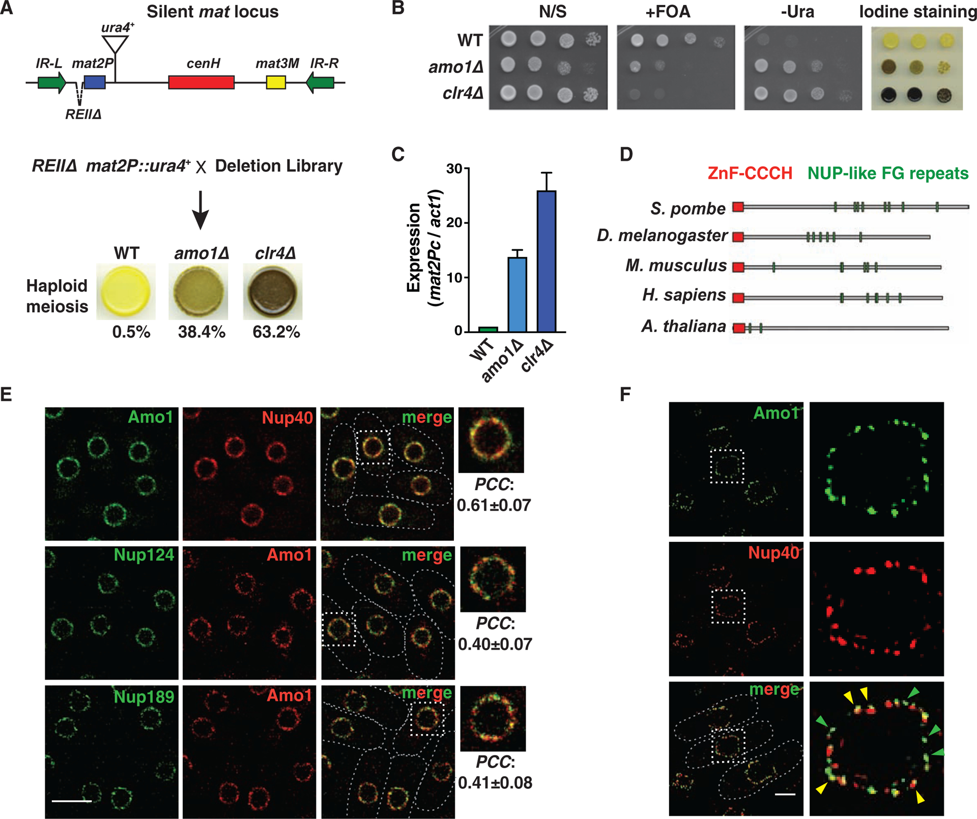
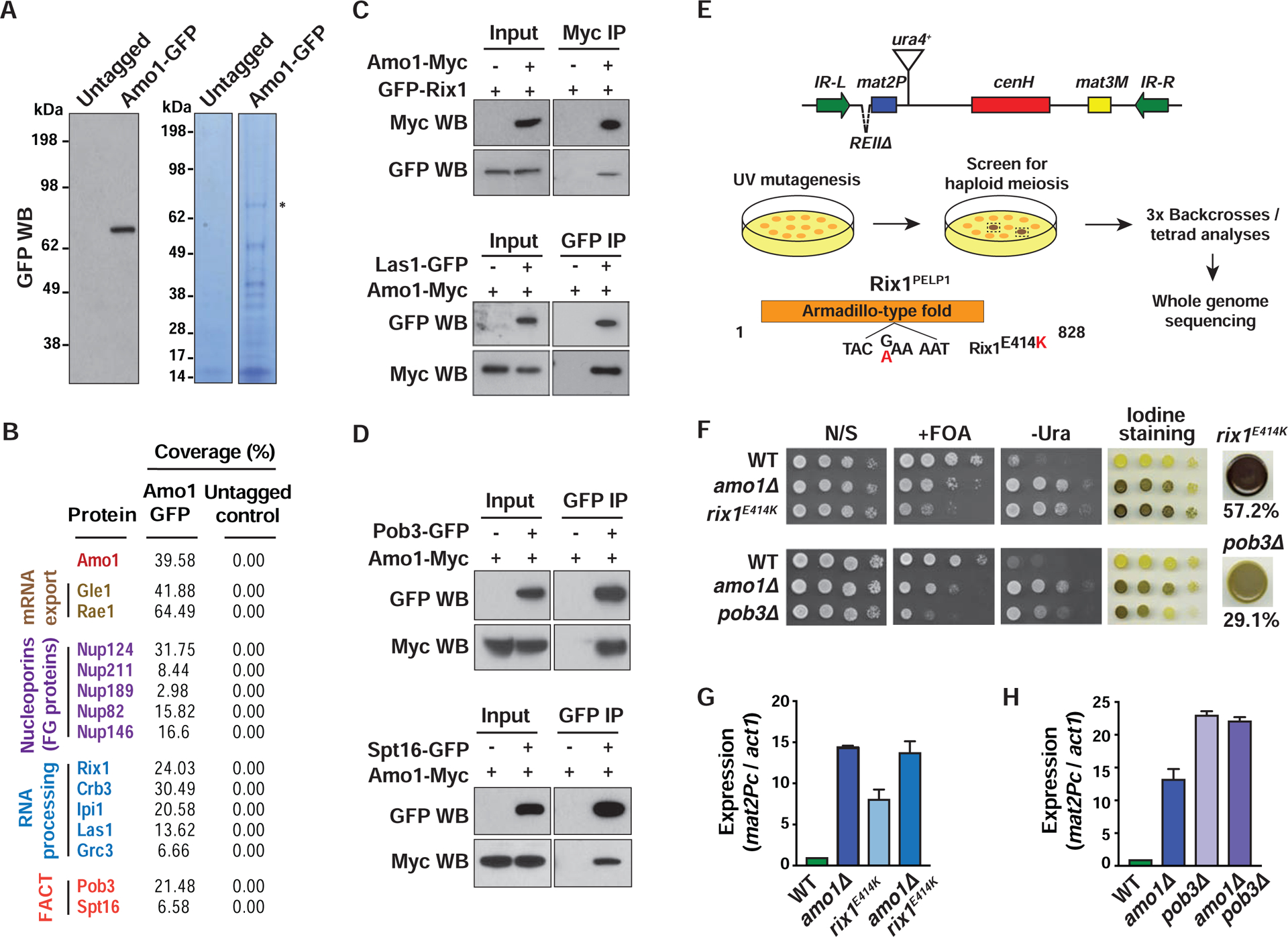
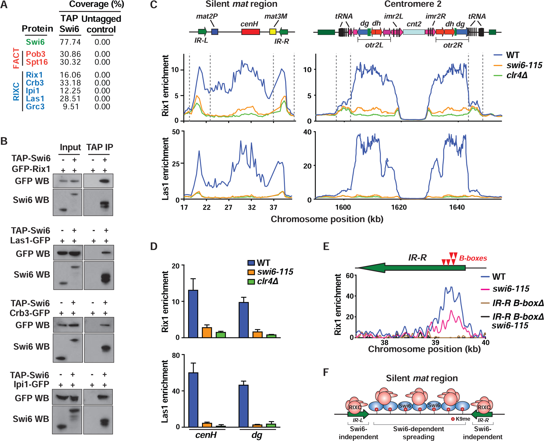
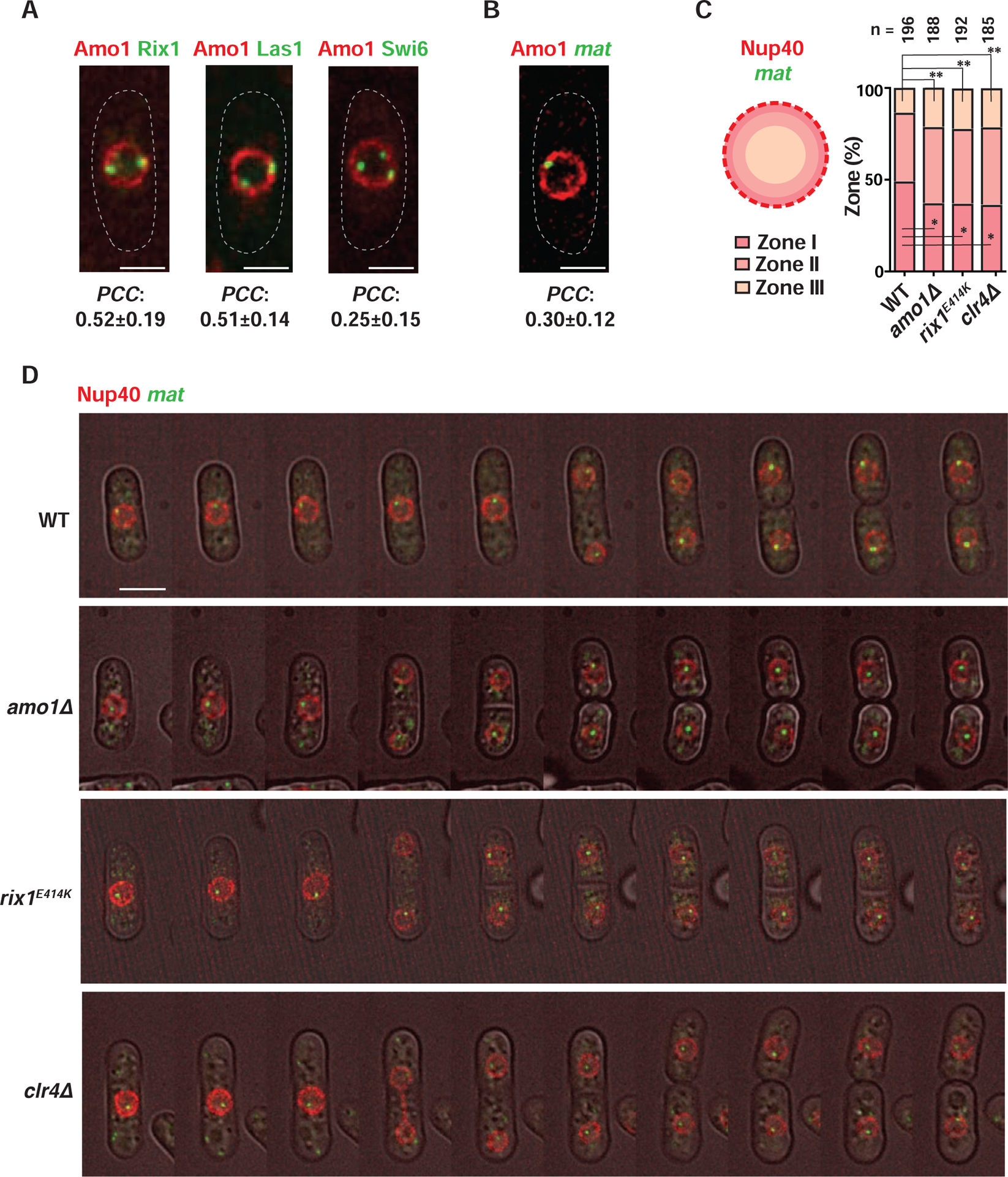
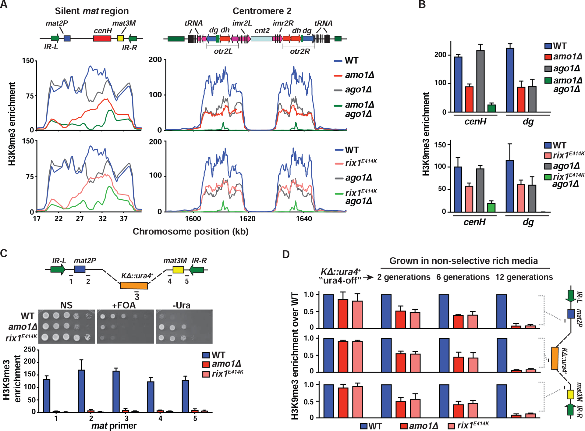
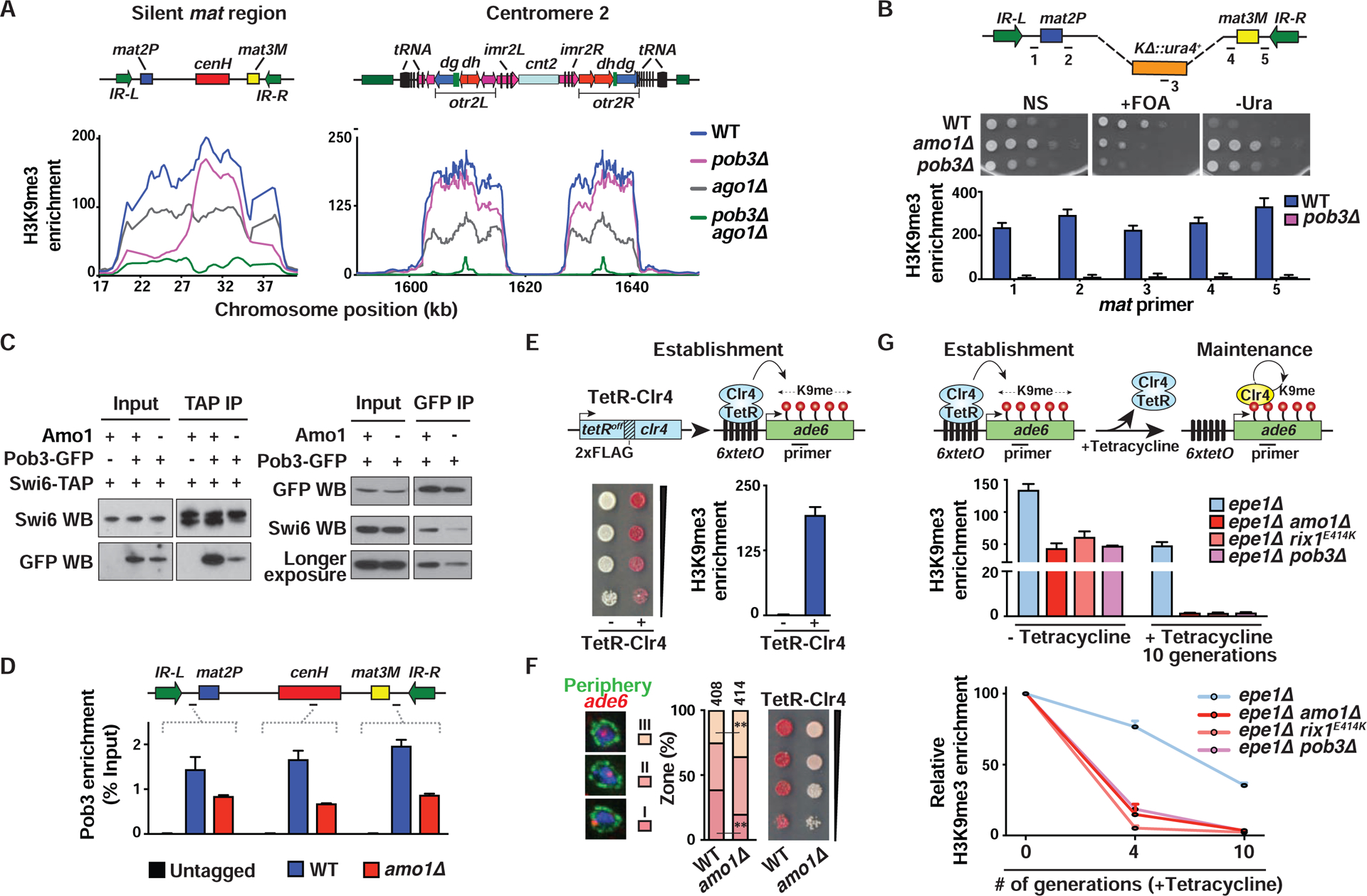
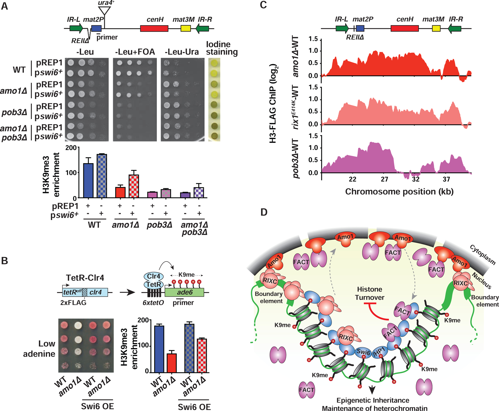
In eukaryotes, heterochromatin is generally located at the nuclear periphery. This study investigates the biological significance of perinuclear positioning for heterochromatin maintenance and gene silencing. We identify the nuclear rim protein Amo1NUPL2 as a factor required for the propagation of heterochromatin at endogenous and ectopic sites in the fission yeast genome. Amo1 associates with the Rix1PELP1-containing RNA processing complex RIXC and with the histone chaperone complex FACT. RIXC, which binds to heterochromatin protein Swi6HP1 across silenced chromosomal domains and to surrounding boundary elements, connects heterochromatin with Amo1 at the nuclear periphery. In turn, the Amo1-enriched subdomain is critical for Swi6 association with FACT that precludes histone turnover to promote gene silencing and preserve epigenetic stability of heterochromatin. In addition to uncovering conserved factors required for perinuclear positioning of heterochromatin, these analyses elucidate a mechanism by which a peripheral subdomain enforces stable gene repression and maintains heterochromatin in a heritable manner.

Keywords: chromatin; epigenetic inheritance; gene regulation; heterochromatin; heterochromatin spreading; histone methylation; nuclear organization; nuclear periphery.


Conflict of interest statement

Declaration of Interests

The authors declare no competing interests.

Figures

Figure 1.
Figure 1.. The nuclear rim protein Amo1 affects heterochromatic silencing.
(A) Genetic screen for factors that affect silencing of the mat locus. Cells grown on minimal media were stained with iodine vapor to detect haploid meiosis. Cells (%) that formed spores in the indicated strains is shown (n>360). (B) Tenfold serial dilution assay of the indicated strains to measure de-repression of mat2P::ura4+. (C) RT-qPCR analysis of mat2Pc expression in the indicated strains (mean ± SD, n=3). (D) Schematic showing the protein domain architecture of Amo1 orthologs. (E) Deconvolution fluorescence imaging of live cells co-expressing GFP- or mCherry-tagged Nups. Insets show magnification of the boxed regions. Co-localization was quantified by Pearson’s correlation coefficient (PCC) (mean ± SD, n>75). (F) Representative structured illumination microscopy (SIM) images. Yellow arrows indicate regions of co-localization, and green arrows show independent Amo1 puncta. See also Figure S1.
Figure 2.
Figure 2.. Amo1 interacts with RIXC and FACT to silence heterochromatin.
(A) Amo1 purification. Western blot confirming Amo1-GFP expression (left). Immunopurified fractions were visualized by Coomassie staining (right). (B) Identities and the total peptide coverage (%) of the Amo1-interacting proteins as determined by mass spectrometry. (C and D) Co-IP of RIXC and FACT subunits with Amo1 in the indicated strains. (E) Genetic screen to identify factors required for heterochromatic silencing. The domain structure of Rix1 with the E414K sequence substitution is shown. (F) Tenfold serial dilution assay of the indicated strains to assess the de-repression of mat2P::ura4+ and detect haploid meiosis. Cells (%) that formed spores in rix1E414K or pob3Δ strains is shown (n>410). (G and H) RT-qPCR analysis of mat2Pc expression in the indicated strains (mean ± SD, n=2). See also Figure S2.
Figure 3.
Figure 3.. RIXC localizes across heterochromatin domains and at boundary elements.
(A) Immunopurified fractions from the indicated strains were subjected to mass spectrometry. The total peptide coverage (%) for the identified proteins is shown. (B) Co-IP of GFP-tagged RIXC with TAP-Swi6. Anti-Swi6 antibody was used to detect endogenous Swi6 (Input, lane 1), TAP-tagged (Input, lane 2) and TEV-cleaved TAP-Swi6 (IP lanes). (C and D) ChIP-chip (C) and ChIP-qPCR (mean ± SD, n=3) (D) showing enrichment of GFP-Rix1 and Las1-GFP at heterochromatic loci. (E) Schematic (top) indicating the positions of B-boxes (red arrows) in the IR-R element. ChIP-seq analysis of GFP-Rix1 in the indicated strains. These strains harbor IR-LΔ to assess IR-R-specific enrichments. The B-boxΔ cells lack a 240-bp sequence that contains 5 B-boxes. (F) Schematic illustrating Swi6-independent and -dependent recruitment of RIXC at mat. See also Figure S3.
Figure 4.
Figure 4.. Amo1 affects the subnuclear localization of the mat locus.
(A and B) Representative fluorescence images of live cells co-expressing Amo1-mCherry and the indicated GFP-tagged proteins (A) or IR-R>lacO/LacI-GFP (mat) (B). Co-localization was quantified by measuring PCC in cells where Rix1, Las1, Swi6 or mat was found at the nuclear periphery (mean ± SD, n>50). (C) Schematic showing localization of mat in different nuclear zones (left) in WT and mutant cells harboring IR-R>lacO/LacI-GFP. Nup40-mCherry marks the NE. Percentage distribution of mat in each frame is counted and plotted for a total of “n” cells. *P<0.05 and **P<0.01 as determined by Student’s t-test for mat in zones I and III respectively (WT vs each mutant). (D) Time lapse images obtained at 20-min intervals are shown for the indicated strains. See also Figure S4 and Videos S1–S4.
Figure 5.
Figure 5.. Amo1 and RIXC affect the epigenetic maintenance of heterochromatin.
(A and B) ChIP-chip (A) and ChIP-qPCR (mean ± SD, n=3) (B) analysis of H3K9me3 enrichments at heterochromatic regions. Note: WT and ago1Δ graphs are the same in the top and bottom panels of (A) as the experiments were performed at the same time. (C) Tenfold serial dilution assay to measure the de-repression of a ura4+ reporter that replaced the K region (KΔ::ura4+) (top). H3K9me3 analysis by ChIP-qPCR (bottom). Fold enrichment values of the target loci (marked in the schematic) are shown as the mean ± SD (n=3). (D) WT and mutant ura4-off strains collected from counter-selective FOA media were grown in non-selective rich media for 2, 6, and 12 generations. ChIP-qPCR showing H3K9me3 enrichment at the indicated loci (see schematic) (mean ± SD, n=2). See also Figure S5.
Figure 6.
Figure 6.. Propagation of endogenous and ectopic heterochromatin domains by Amo1-RIXC and FACT.
(A) H3K9me3 ChIP-chip at the heterochromatic regions. (B) Tenfold serial dilution (top) and H3K9me3 enrichments (bottom, mean ± SD, n=2) are shown for the indicated strains. (C) Co-IPs were performed using anti-GFP agarose beads (left) or anti-TAP beads (right) followed by western blotting. (D) ChIP-qPCR showing Pob3-GFP enrichments. Percent Input values of the target loci are shown as the mean ± SD (n=2). (E) Serial dilutions on low adenine EMM medium and ChIP-qPCR analysis of H3K9me3 (mean ± SD, n=2) in the indicated TetR-Clr4-expressing 6xtetO-ade6+ reporter strains. (F) Representative images of ade6+ DNA FISH in the three zones (left). ade6+ locus (%) located in each zone (“n” cells from two independent experiments) (middle). **P<0.01 as determined by Student’s t-test for ade6+ locus in zones III and I (WT vs amo1Δ). Serial dilutions of the indicated strains (right). (G) Establishment and maintenance of heterochromatin at an ectopic site. Endogenous Clr4 is indicated in yellow (top). H3K9me3 ChIP-qPCR in strains grown in EMM+tetracycline media for 0, 4, and 10 generations (mean ± SD, n=2). Fold enrichments were calculated relative to vps33 (middle) or relative to “-tetracycline” (0 generations) (bottom). See also Figure S6.
Figure 7.
Figure 7.. Nuclear peripheral positioning of heterochromatin by Amo1 suppresses histone turnover.
(A and B) Tenfold serial dilution and ChIP-qPCR analysis of H3K9me3 enrichments (mean ± SD, n=2) in WT and mutant strains transformed with LEU2-based plasmids pREP1 (control) or pswi6+ (A), or the indicated TetR-Clr4-expressing 6xtetO-ade6+ reporter strains (B). (C) Histone turnover at the mat locus as determined by H3-FLAG ChIP-chip. Strains contained REIIΔ that permits modifications affecting heterochromatin maintenance to be easily detected (see also STAR★Methods). The signals were normalized to WT. (D) Model: Amo1 positions heterochromatic loci at the nuclear periphery via RIXC and facilitates efficient loading of FACT onto Swi6HP1-bound heterochromatin. Together with HDACs (not shown), they promote epigenetic inheritance and heterochromatin maintenance by suppressing histone turnover. See also Figure S7.

References

    1. Akhtar A, and Gasser SM (2007). The nuclear envelope and transcriptional control. Nat. Rev. Genet 8, 507–517. - PubMed
    1. Al-Sady B, Madhani HD, and Narlikar GJ (2013). Division of labor between the chromodomains of HP1 and Suv39 methylase enables coordination of heterochromatin spread. Mol. Cell 51, 80–91. - PMC - PubMed
    1. Alfredsson-Timmins J, Henningson F, and Bjerling P (2007). The Clr4 methyltransferase determines the subnuclear localization of the mating-type region in fission yeast. J. Cell Sci 120, 1935–1943. - PubMed
    1. Andrulis ED, Neiman AM, Zappulla DC, and Sternglanz R (1998). Perinuclear localization of chromatin facilitates transcriptional silencing. Nature 394, 592–595. - PubMed
    1. Asakawa H, Yang HJ, Yamamoto TG, Ohtsuki C, Chikashige Y, Sakata-Sogawa K, Tokunaga M, Iwamoto M, Hiraoka Y, and Haraguchi T (2014). Characterization of nuclear pore complex components in fission yeast Schizosaccharomyces pombe. Nucleus 5, 149–162. - PMC - PubMed

Publication types

MeSH terms