

Rare squamous cell carcinoma arising from a presacral epidermoid cyst: A case report
- PMID: 31884265
- PMCID: PMC6939061
- DOI: 10.1016/j.ijscr.2019.12.022
Rare squamous cell carcinoma arising from a presacral epidermoid cyst: A case report
Abstract
Introduction: Presacral epidermoid cysts are uncommon, usually benign cysts caused by developmental abnormalities in the fetal period. We present a rare case of squamous cell carcinoma arising from a presacral epidermoid cyst.
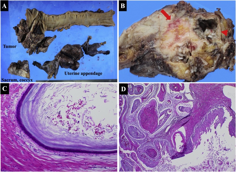
Presentation of case: A 59-year-old woman complained of tenesmus and discomfort in the buttocks. Computed tomography revealed a 50-mm well-defined cystic mass in the presacrum and a 70-mm solid mass extending from the cyst into the rectum, vagina, and left sciatic spine. On T1-weighted magnetic resonance images, the cyst was unilocular and the mass was marginated with low intensity. On T2-weighted images, the mass had high intensity. A malignant presacral developmental cyst was diagnosed, without obvious metastasis. Using abdominal and parasacral approaches, Hartmann's operation was performed with multiorgan resection, including the sacrum, coccyx, left sciatic spine, internal obturator muscle, rectum, and uterine appendage. Histopathology of the excised specimen revealed a squamous cell carcinoma originating from the presacral epidermoid cyst.
Discussion: Reports of malignant transformation of epidermoid cysts in the presacral space, as in the present case, are extremely rare. Because of their unusual location and slow growth, epidermoid cysts tend to remain asymptomatic. Because the patient had a malignant tumor with suspected invasion of adjacent organs, combination surgery was selected.
Conclusion: Although further research is required, presacral epidermoid cysts are extremely rare and may be malignant. Thorough preoperative image evaluation is crucial for complete resection.
Keywords: Benign cysts; Fetal period; Presacral epidermoid cysts; Squamous cell carcinoma.
Copyright © 2019 The Author(s). Published by Elsevier Ltd.. All rights reserved.
Conflict of interest statement
Declaration of Competing Interest The authors declare no conflicts of interest.
Figures


Similar articles
-
A Case of Oropharyngeal Carcinoma Accompanying a Presacral Malignant Epidermoid Cyst.Cureus. 2024 Sep 21;16(9):e69841. doi: 10.7759/cureus.69841. eCollection 2024 Sep. Cureus. 2024. PMID: 39435196 Free PMC article.
-
A report of presacral epidermoid cyst in perimenopausal women: An extremely rare site and an unusual cause of chronic constipation.Int J Surg Case Rep. 2023 Feb;103:107880. doi: 10.1016/j.ijscr.2023.107880. Epub 2023 Jan 9. Int J Surg Case Rep. 2023. PMID: 36634501 Free PMC article.
-
Laparoscopic surgery of a presacral epidermoid cyst: A case report.Int J Surg Case Rep. 2019;59:23-26. doi: 10.1016/j.ijscr.2019.04.043. Epub 2019 May 4. Int J Surg Case Rep. 2019. PMID: 31102835 Free PMC article.
-
A Rare Transformation of Epidermoid Cyst into Squamous Cell Carcinoma: A Case Report with Literature Review.Am J Case Rep. 2019 Aug 3;20:1141-1143. doi: 10.12659/AJCR.912828. Am J Case Rep. 2019. PMID: 31375657 Free PMC article. Review.
-
Presacral epidermoid cyst in a male: a case report and literature review.J Surg Educ. 2010 Jul-Aug;67(4):227-32. doi: 10.1016/j.jsurg.2010.06.005. J Surg Educ. 2010. PMID: 20816358 Review.
Cited by
-
Epidermoid Cyst of the Cecum Treated by Laparoscopic Colectomy: A Case Report With Histopathology and Literature Review.Case Rep Gastrointest Med. 2025 Jun 13;2025:6326844. doi: 10.1155/crgm/6326844. eCollection 2025. Case Rep Gastrointest Med. 2025. PMID: 40546654 Free PMC article.
-
A Case of Oropharyngeal Carcinoma Accompanying a Presacral Malignant Epidermoid Cyst.Cureus. 2024 Sep 21;16(9):e69841. doi: 10.7759/cureus.69841. eCollection 2024 Sep. Cureus. 2024. PMID: 39435196 Free PMC article.
-
Malignant transformation of tailgut cyst to squamous cell carcinoma, a rare case with poor outcome.Clin Case Rep. 2023 Feb 5;11(2):e6893. doi: 10.1002/ccr3.6893. eCollection 2023 Feb. Clin Case Rep. 2023. PMID: 36777790 Free PMC article.
-
Epidermoid cyst of the cecum resected by single-incision laparoscopic colectomy: a case report.Surg Case Rep. 2021 Feb 25;7(1):57. doi: 10.1186/s40792-021-01138-2. Surg Case Rep. 2021. PMID: 33630182 Free PMC article.
References
-
- Agha R.A., Borrelli M.R., Farwana R., Koshy K., Fowler A., Orgill D.P., For the SCARE Group The SCARE 2018 statement: updating consensus Surgical CAse REport (SCARE) guidelines. Int. J. Surg. 2018;60:132–136. - PubMed
-
- Hawkins W.J., Jackman R.J. Developmental cysts as a source of perianal abscesses, sinuses and fistulas. Am. J. Surg. 1953;86:678–683. - PubMed
-
- Yang D.M., Kim H.C., Lee H.L., Lee S.H., Kim G.Y. Squamous cell carcinoma arising from a presacral epidermoid cyst: CT and MR findings. Abdom. Imaging. 2008;33:498–500. - PubMed
Publication types
LinkOut - more resources
Full Text Sources
Research Materials
