. 2020 Feb 17;375(1792):20190164.
doi: 10.1098/rstb.2019.0164. Epub 2019 Dec 30.

The unity and diversity of the ciliary central apparatus

Affiliations

The unity and diversity of the ciliary central apparatus

Lei Zhao et al. Philos Trans R Soc Lond B Biol Sci. .

Abstract

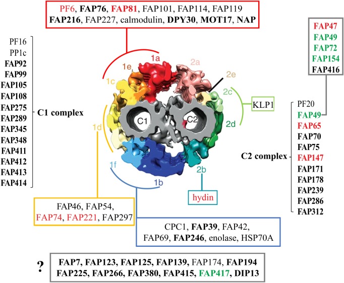
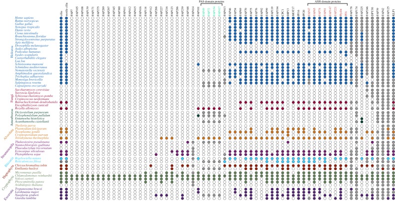
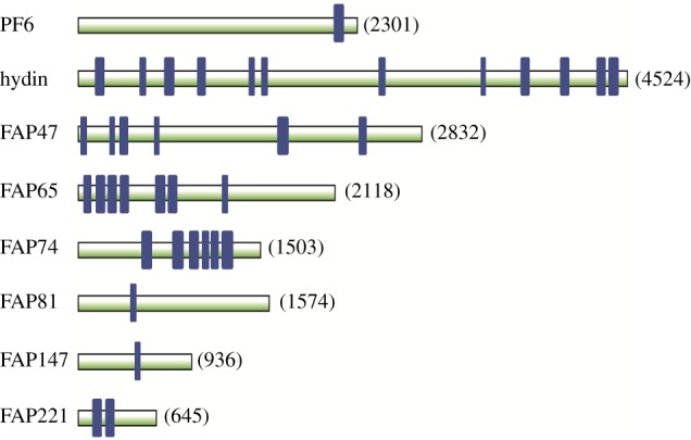
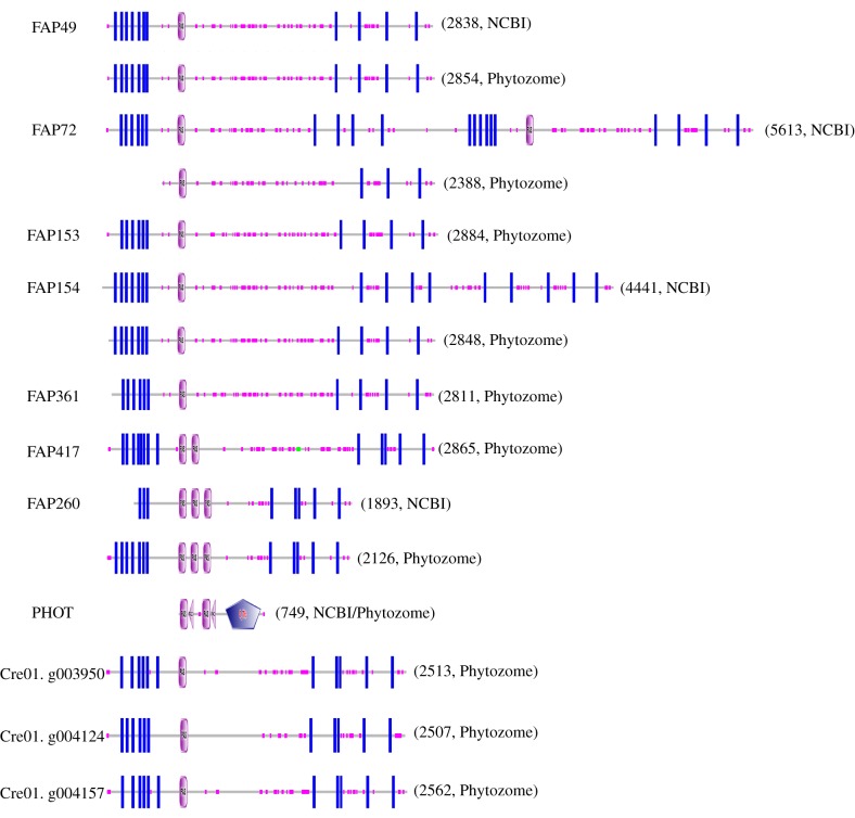
Nearly all motile cilia and flagella (terms here used interchangeably) have a '9+2' axoneme containing nine outer doublet microtubules and two central microtubules. The central pair of microtubules plus associated projections, termed the central apparatus (CA), is involved in the control of flagellar motility and is essential for the normal movement of '9+2' cilia. Research using the green alga Chlamydomonas reinhardtii, an important model system for studying cilia, has provided most of our knowledge of the protein composition of the CA, and recent work using this organism has expanded the number of known and candidate CA proteins nearly threefold. Here we take advantage of this enhanced proteome to examine the genomes of a wide range of eukaryotic organisms, representing all of the major phylogenetic groups, to identify predicted orthologues of the C. reinhardtii CA proteins and explore how widely the proteins are conserved and whether there are patterns to this conservation. We also discuss in detail two contrasting groups of CA proteins-the ASH-domain proteins, which are broadly conserved, and the PAS proteins, which are restricted primarily to the volvocalean algae. This article is part of the Theo Murphy meeting issue 'Unity and diversity of cilia in locomotion and transport'.

Keywords: ASH-domain proteins; Chlamydomonas; PAS-domain proteins; axoneme evolution; central microtubules; flagella.


Conflict of interest statement

The authors declare no competing financial interests.

Figures

Figure 1.
Figure 1.
Summary of CA proteins and their predicted locations in the C1 and C2 microtubules. Diagram of cross-section of the Chlamydomonas reinhardtii CA (modified from [1]) showing predicted locations of CA proteins including novel candidate or confirmed CA proteins identified by Zhao et al. [2] (bold font); FAP76 and FAP216 were localized by Fu et al. [3]. ‘1a’–‘1f’ and ‘2a’–‘2e’ indicate projections C1a to C1f and C2a to C2e, respectively. ASH-domain proteins are in red font; PAS-domain proteins are in green font. Some proteins are predicted to be associated with either the C1 or C2 microtubule, but their specific locations are not yet determined; others (red, green, turquoise, dark blue and yellow boxes) are predicted to be associated with specific projections, pairs of projections or a supercomplex consisting of the C1a, C1e and C1c projections. The FAP47 complex (box, upper right) is likely to be associated with C2 based on solubility properties of FAP49. The question mark indicates proteins whose locations in the CA are not yet known. Modified from Zhao et al. [2]. (Online version in colour.)
Figure 2.
Figure 2.
Dot plot showing phylogenetic distribution of orthologues of C. reinhardtii CA-specific proteins. Species belonging to the same major taxonomic group are indicated by text with the same colour. Coloured dots indicate either the presence of cilia or motile cilia (first and second columns, respectively) or the presence of at least one likely orthologue in the indicated species. Empty dots indicate the absence of cilia, motile cilia or orthologues. Grey dots for Reticulomyxa filosa indicate uncertainty with regard to the presence of cilia (see text). Hatched dots indicate that sequences from these species were present in the OrthoFinder gene tree but eliminated as likely orthologues because they were in a different clade from the C. reinhardtii query protein (see Methods). Proteins most affected in this way were members of large families or contained common conserved motifs and included KLP1 and FAP125 (kinesin superfamily), FAP42 (transferase family), FAP39 (ATPase), DPY30 (DPY-30 domain), FAP246 (LRR repeats and EF-hands), FAP225 (EF-hands), FAP174 (MYC-binding) and FAP178 (calponin domain). PAS- and ASH-domain proteins (green and red font, respectively) are indicated. (Online version in colour.)
Figure 3.
Figure 3.
Predicted ASH domains of C. reinhardtii CA proteins. ASH domains are indicated by blue rectangles. Proteins are drawn to scale; the number to the right of each protein indicates the number of amino acids in the protein. ASH domains were identified as described in the Methods. (Online version in colour.)
Figure 4.
Figure 4.
Domain architecture of predicted C. reinhardtii flagellar proteins containing PAS domains. Pink ovals marked ‘PAS’ indicate predicted PAS domains. Blue rectangles indicate predicted transmembrane domains. Small pink boxes along each protein indicate regions of low sequence complexity. In PHOT, pink triangles marked ‘PAC’ indicate ‘motif C-terminal to PAS motifs (likely to contribute to PAS structural domain)’ and the blue pentagon marked ‘S_TKc’ indicates a ‘serine/threonine protein kinases, catalytic domain’. The number to the right of each architecture indicates the number of amino acids in the protein sequence. PAS domains were identified as described in the Methods. When there is a discrepancy between sequences from NCBI and Phytozome, predictions for both sequences are shown. The locations of exclusive unique peptides suggested that some Phytozome models were more accurate than the corresponding NCBI models; e.g. for FAP72, unique peptides were predicted to originate from throughout the N-terminal half of the NCBI model (most of which is identical to the Phytozome model) but not the C-terminal half. (Online version in colour.)
Figure 5.
Figure 5.
Maximum-likelihood tree of C. reinhardtii PAS proteins and orthologues. Maximum-likelihood tree of C. reinhardtii flagellar-associated PAS proteins with high-confidence orthologues, rooted with R. allomycis as the outgroup. Green highlighting indicates C. reinhardtii PAS proteins predicted to be associated with the CA; orange highlighting indicates C. reinhardtii flagellar PAS proteins encoded on Chromosome 1; blue highlighting indicates other C. reinhardtii flagellar PAS proteins. For the tree construction only, a model for FAP72 was used that included the N-terminus of NCBI FAP72 (aa 1–471) joined to the N-terminus of Phytozome FAP72 (see Methods). The conjoined model lacks only 15 unmatched amino acids from the C-terminus of Phytozome FAP408. Phytozyme lists two transcripts for the Cre01.g004124 gene: Cre01.g004124.t1.1 (primary) and Cre01.g004124.t2.1; the former includes two amino acids not present in the latter and was used to construct this tree, whereas the latter is the one associated with this gene in the Chlamydomonas Flagellar Proteome Project (http://chlamyfp.org/index.php) and so is referred to in the main text. The tree was constructed with IQ-TREE [31] and was visualized using iTOL [34]. Support values show ultrafast bootstrap data with 1000 iterations [33]. Scale represents average number of substitutions per residue. (Online version in colour.)

References

    1. Carbajal-Gonzalez BI, Heuser T, Fu X, Lin J, Smith BW, Mitchell DR, Nicastro D. 2013. Conserved structural motifs in the central pair complex of eukaryotic flagella. Cytoskeleton 70, 101–120. (10.1002/cm.21094) - DOI - PMC - PubMed
    1. Zhao L, Hou Y, Picariello T, Craige B, Witman GB. 2019. Proteome of the central apparatus of a ciliary axoneme. J. Cell Biol. 218, 2051–2070. (10.1083/jcb.201902017) - DOI - PMC - PubMed
    1. Fu G, et al. 2019. Structural organization of the C1a-e-c supercomplex within the ciliary central apparatus. J. Cell Biol. 218, 4236–4251. (10.1083/jcb.201906006). - DOI - PMC - PubMed
    1. Nakazawa Y, Ariyoshi T, Noga A, Kamiya R, Hirono M. 2014. Space-dependent formation of central pair microtubules and their interactions with radial spokes. PLoS ONE 9, e110513 (10.1371/journal.pone.0110513) - DOI - PMC - PubMed
    1. Mitchell DR. 2017. Evolution of cilia. Cold Spring Harb. Perspect. Biol. 9, a028290 (10.1101/cshperspect.a028290) - DOI - PMC - PubMed

Publication types

MeSH terms